PDGFR alpha Recombinant Rabbit Monoclonal Antibody [JF104-6]


Catalog# ET1702-49
PDGFR alpha Recombinant Rabbit Monoclonal Antibody [JF104-6]
-
WB
-
IHC-Fr
-
Human
-
Mouse
-
Rat
-
Cynomolgus monkey
-
Pig
-
unconjugated
概述
产品名称
PDGFR alpha Recombinant Rabbit Monoclonal Antibody [JF104-6]
抗体类型
Recombinant Rabbit monoclonal Antibody
免疫原
Synthetic peptide within C-terminal human PDGFR alpha.
种属反应性
Human, Mouse, Rat (Predicted: Cynomolgus monkey, Pig)
验证应用
WB, IHC-Fr
分子量
Predicted band size: 123 kDa
阳性对照
Mouse embryonic femur tissue, NIH/3T3 cell lysates.
偶联
unconjugated
克隆号
JF104-6
RRID
产品特性
形态
Liquid
浓度
存放说明
Shipped at 4℃. Store at +4℃ short term (1-2 weeks). It is recommended to aliquot into single-use upon delivery. Store at -20℃ long term.
存储缓冲液
1*TBS (pH7.4), 0.05% BSA, 40% Glycerol. Preservative: 0.05% Sodium Azide.
亚型
IgG
纯化方式
Protein A affinity purified.
应用稀释度
-
WB
-
1:5,000-1:10,000
-
IHC-Fr
-
1:500
靶点
功能
Platelet-derived growth factor (PDGF) is a mitogen for mesenchyme- and glia-derived cells. PDGF consists of two chains, A and B, which dimerize to form functionally distinct isoforms, PGDF-AA, PDGF-AB and PDGF-BB. These three isoforms bind with different affinities to two receptor types, PDGFR-α and -β, which are endowed with protein tyrosine kinase domains. PDGFR-α can bind to both A and B subunits of PDGF, while PDGFR-β can only bind the B subunit. Ligand binding promotes either homo- or heterodimerization of the PDGF receptors in a specific manner. PDGF-AA induces the dimerization of two α receptors, PDGF-AB induces dimerization of αα and αβ and PDGF-BB induces the formation of three types of dimers, αα, αβ and ββ. Translocation of the PDGFR-β gene with the Tel gene is linked to chronic myelomonocytic leukemia (CMML), a myelodysplastic syndrome, and demonstrates the oncogenic potential of the PDGF receptors.
背景文献
1. Benedykcinska A et al. Generation of brain tumours in mice by Cre-mediated recombination of neural progenitors in situ with the tamoxifen metabolite endoxifen. Dis Model Mech 9:211-20 (2016).
2. Rondahl V et al. Lrig2-deficient mice are protected against PDGFB-induced glioma. PLoS One 8:e73635 (2013).
序列相似性
Belongs to the protein kinase superfamily. Tyr protein kinase family. CSF-1/PDGF receptor subfamily.
组织特异性
Detected in platelets (at protein level). Widely expressed. Detected in brain, fibroblasts, smooth muscle, heart, and embryo. Expressed in primary and metastatic colon tumors and in normal colon tissue.
翻译后修饰
N-glycosylated.; Ubiquitinated, leading to its internalization and degradation.; Autophosphorylated on tyrosine residues upon ligand binding. Autophosphorylation occurs in trans, i.e. one subunit of the dimeric receptor phosphorylates tyrosine residues on the other subunit. Phosphorylation at Tyr-731 and Tyr-742 is important for interaction with PIK3R1. Phosphorylation at Tyr-720 and Tyr-754 is important for interaction with PTPN11. Phosphorylation at Tyr-762 is important for interaction with CRK. Phosphorylation at Tyr-572 and Tyr-574 is important for interaction with SRC and SRC family members. Phosphorylation at Tyr-988 and Tyr-1018 is important for interaction with PLCG1.
亚细胞定位
Golgi apparatus, Cell membrane, cilium.
别名
Alpha-type platelet-derived growth factor receptor antibody
CD140 antigen-like family member A antibody
CD140a antibody
CD140a antigen antibody
MGC74795 antibody
PDGF alpha chain antibody
PDGF-R-alpha antibody
PDGFR 2 antibody
PDGFR alpha antibody
PDGFR2 antibody
展开图片
-
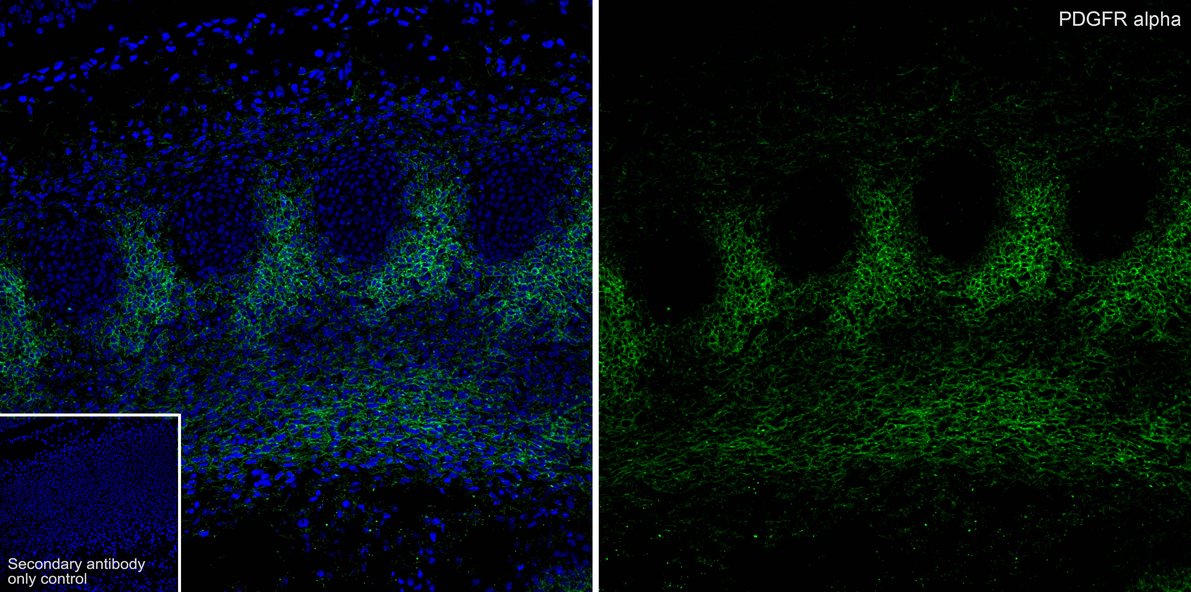
Application: IHC-Fr
Species: Mouse
Site: E14.5 embyro
Sample: Frozen section
Antibody concentration: 1:500
Antigen retrieval: Not required -
Western blot analysis of PDGFR alpha on NIH/3T3 cell lysates with Rabbit anti-PDGFR alpha antibody (ET1702-49) at 1/5,000 dilution.
Lysates/proteins at 10 µg/Lane.
Predicted band size: 123 kDa
Observed band size: 180 kDa
Exposure time: 2 minutes;
6% SDS-PAGE gel.
Proteins were transferred to a PVDF membrane and blocked with 5% NFDM/TBST for 1 hour at room temperature. The primary antibody (ET1702-49) at 1/5,000 dilution was used in 5% NFDM/TBST at room temperature for 2 hours. Goat Anti-Rabbit IgG - HRP Secondary Antibody (HA1001) at 1/50,000 dilution was used for 1 hour at room temperature. -
☑ Knockdown (KD)
Western blot analysis of PDGFR alpha on different lysates with Rabbit anti-PDGFR alpha antibody (ET1702-49) at 1/5,000 dilution.
Lane 1: NIH/3T3-si NT cell lysate
Lane 2: NIH/3T3-si PDGFR alpha cell lysate
Lysates/proteins at 10 µg/Lane.
Predicted band size: 123 kDa
Observed band size: 190 kDa
Exposure time: 2 minutes;
4-20% SDS-PAGE gel.
Proteins were transferred to a PVDF membrane and blocked with 5% NFDM in TBST for 1 hour at room temperature. The primary antibody (ET1702-49, 1/5,000) and Loading control antibody (Rabbit anti-GAPDH, ET1601-4, 1/10,000) were used in 5% BSA at room temperature for 2 hours. Goat Anti-rabbit IgG-HRP Secondary Antibody (HA1001) at 1/50,000 dilution was used for 1 hour at room temperature.
请注意: All products are "FOR RESEARCH USE ONLY AND ARE NOT INTENDED FOR DIAGNOSTIC OR THERAPEUTIC USE"
引文
-
Rebamipide Induces Hair Regeneration Through EP4-Driven Lipid Metabolism Remodeling
期刊: International Journal Of Molecular Sciences
DOI: 10.3390/ijms262010132
IF: 4.9
应用: IF-Tissue,WB
反应种属: Mouse
发表时间: 2025 Oct
-
Aldh1L1 Lineage Cells Contribute to the Functional Heterogeneity Within the Cells in Glial Scars After Spinal Cord Injury Through YAP Signaling
期刊: Glia
DOI: 10.1002/glia.70092
IF: 5.1
应用: IF-Tissue
反应种属: Mouse
发表时间: 2025 Oct
-
PPY-Induced iCAFs Cultivate an Immunosuppressive Microenvironment in Pancreatic Cancer
期刊: Advanced Science
DOI: 10.1002/advs.202413432
IF: 14.3
应用: mIHC
反应种属: Human,Mouse
发表时间: 2025 Mar
-
Cavity oscillation drives pattern formation in early mammalian embryos
期刊: Cell Reports
DOI: 10.1016/j.celrep.2025.115342
IF: 7.5
应用: IF
反应种属: Mouse
发表时间: 2025 Feb
-
Crosstalk between cancer-associated fibroblasts and myeloid cells shape the heterogeneous microenvironment of gastric cancer
期刊: Current Genomics
DOI:
IF: 1.8
应用: IHC-P
反应种属: Human
发表时间: 2024 Jul
-
Oncogenic RTKs sensitize cancer cells to ferroptosis via c-Myc mediated upregulation of ACSL4
期刊: Cell Death & Disease
DOI:
IF: 8.1
应用: WB
反应种属: Human
发表时间: 2024 Dec
-
Establishment and characterization of a skeletal myoblast cell line of grass carp (Ctenopharyngodon idellus)
期刊: Fish Physiology And Biochemistry
DOI: 10.1007/s10695-023-01246-w
IF: 2.9
应用: WB,IF
反应种属: Grass carp,Zebrafish,Mouse
发表时间: 2023 Oct
-
Spatial transcriptomics atlas reveals the crosstalk between cancer-associated fibroblasts and tumor microenvironment components in colorectal cancer
期刊: Journal Of Translational Medicine
DOI:
IF: 8.440
应用: IF,IHC-P
反应种属: Human
发表时间: 2022 Jul
 浙公网安备 33019202000643号