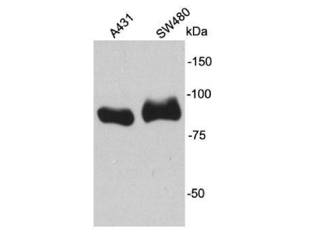
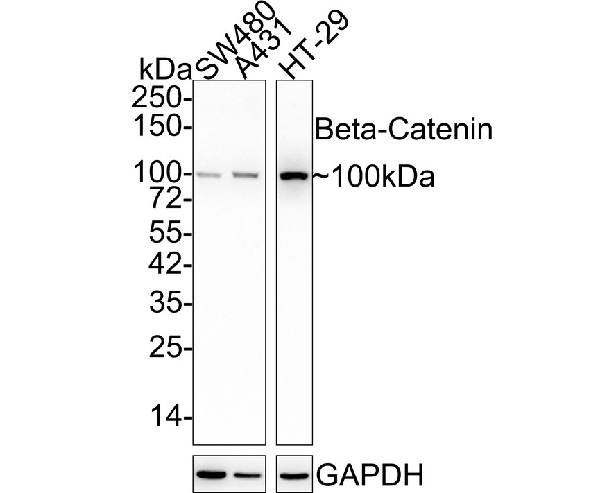
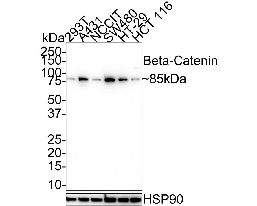
概述
产品名称
Beta Catenin Rabbit Polyclonal Antibody
抗体类型
Rabbit Polyclonal Antibody
免疫原
Synthetic peptide within C-terminal human Beta-Catenin.
种属反应性
Human, Mouse, Rat
验证应用
WB, IHC-P, FC, IF-Cell, IF-Tissue
分子量
Predicted band size: 85 kDa
阳性对照
SW480 cell lysate, HT-29 cell lysate, NIH/3T3 cell lysate, C6 cell lysate, Human brain tissue lysate, Mouse brain tissue lysate, Rat brain tissue lysate, HT-29, C6, human breast cancer tissue, human colon cancer tissue, mouse colon tissue, rat colon tissue.
偶联
unconjugated
RRID
产品特性
形态
Liquid
浓度
1ug/ul
存放说明
Store at +4℃ after thawing. Aliquot store at -20℃. Avoid repeated freeze / thaw cycles.
存储缓冲液
1*PBS (pH7.4), 0.2% BSA, 40% Glycerol. Preservative: 0.05% Sodium Azide.
亚型
IgG
纯化方式
Immunogen affinity purified.
应用稀释度
-
WB
-
1:1,000
-
IHC-P
-
1:5,000
-
FC
-
1:1,000
-
IF-Cell
-
1:100-1:500
-
IF-Tissue
-
1:200-1:1,000
发表文章中的应用
发表文章中的种属
| Mouse | See 4 publications below |
| Human | See 2 publications below |
靶点
功能
Key downstream component of the canonical Wnt signaling pathway In the absence of Wnt, forms a complex with AXIN1, AXIN2, APC, CSNK1A1 and GSK3B that promotes phosphorylation on N-terminal Ser and Thr residues and ubiquitination of CTNNB1 via BTRC and its subsequent degradation by the proteasome. In the presence of Wnt ligand, CTNNB1 is not ubiquitinated and accumulates in the nucleus, where it acts as a coactivator for transcription factors of the TCF/LEF family, leading to activate Wnt responsive genes Involved in the regulation of cell adhesion, as component of an E-cadherin:catenin adhesion complex. Acts as a negative regulator of centrosome cohesion. Involved in the CDK2/PTPN6/CTNNB1/CEACAM1 pathway of insulin internalization. Blocks anoikis of malignant kidney and intestinal epithelial cells and promotes their anchorage-independent growth by down-regulating DAPK2. Disrupts PML function and PML-NB formation by inhibiting RANBP2-mediated sumoylation of PML. Promotes neurogenesis by maintaining sympathetic neuroblasts within the cell cycle. Involved in chondrocyte differentiation via interaction with SOX9: SOX9-binding competes with the binding sites of TCF/LEF within CTNNB1, thereby inhibiting the Wnt signaling.
背景文献
1. "Oncogenic beta-catenin is required for bone morphogenetic protein 4 expression in human cancer cells." Kim J.-S., Crooks H., Dracheva T., Nishanian T.G., Singh B., Jen J., Waldman T. Cancer Res. 62:2744-2748(2002)"
2. "Beta-catenin expression in pilomatrixomas. Relationship with beta-catenin gene mutations and comparison with beta-catenin expression in normal hair follicles." Moreno-Bueno G., Gamallo C., Perez-Gallego L., Contreras F., Palacios J. Br. J. Dermatol. 145:576-581(2001)
3. "EBP50, a beta-catenin-associating protein, enhances Wnt signaling and is over-expressed in hepatocellular carcinoma." Shibata T., Chuma M., Kokubu A., Sakamoto M., Hirohashi S. Hepatology 38:178-186(2003)
4. "Identification of beta-catenin as a target of the intracellular tyrosine kinase PTK6." Palka-Hamblin H.L., Gierut J.J., Bie W., Brauer P.M., Zheng Y., Asara J.M., Tyner A.L. J. Cell Sci. 123:236-245(2010)
序列相似性
Belongs to the beta-catenin family.
组织特异性
Expressed in several hair follicle cell types: basal and peripheral matrix cells, and cells of the outer and inner root sheaths. Expressed in colon. Present in cortical neurons (at protein level). Expressed in breast cancer tissues (at protein level).
翻译后修饰
Phosphorylation at Ser-552 by AMPK promotes stabilizion of the protein, enhancing TCF/LEF-mediated transcription (By similarity). Phosphorylation by GSK3B requires prior phosphorylation of Ser-45 by another kinase. Phosphorylation proceeds then from Thr-41 to Ser-37 and Ser-33. Phosphorylated by NEK2. EGF stimulates tyrosine phosphorylation. Phosphorylation on Tyr-654 decreases CDH1 binding and enhances TBP binding. Phosphorylated on Ser-33 and Ser-37 by HIPK2 and GSK3B, this phosphorylation triggers proteasomal degradation. Phosphorylation on Ser-191 and Ser-246 by CDK5. Phosphorylation by CDK2 regulates insulin internalization. Phosphorylation by PTK6 at Tyr-64, Tyr-142, Tyr-331 and/or Tyr-333 with the predominant site at Tyr-64 is not essential for inhibition of transcriptional activity.; Ubiquitinated by the SCF(BTRC) E3 ligase complex when phosphorylated by GSK3B, leading to its degradation. Ubiquitinated by a E3 ubiquitin ligase complex containing UBE2D1, SIAH1, CACYBP/SIP, SKP1, APC and TBL1X, leading to its subsequent proteasomal degradation. Ubiquitinated and degraded following interaction with SOX9 (By similarity).; S-nitrosylation at Cys-619 within adherens junctions promotes VEGF-induced, NO-dependent endothelial cell permeability by disrupting interaction with E-cadherin, thus mediating disassembly adherens junctions.; O-glycosylation at Ser-23 decreases nuclear localization and transcriptional activity, and increases localization to the plasma membrane and interaction with E-cadherin CDH1.; Deacetylated at Lys-49 by SIRT1.
亚细胞定位
Cytoplasm, Nucleus, Cell junction, Cell membrane, Cytoskeleton, Synapse.
别名
β Catenin
Beta catenin antibody
Beta-catenin antibody
Cadherin associated protein antibody
Catenin (cadherin associated protein), beta 1, 88kDa antibody
Catenin beta 1 antibody
Catenin beta-1 antibody
CATNB antibody
CHBCAT antibody
CTNB1_HUMAN antibody
展开β Catenin
Beta catenin antibody
Beta-catenin antibody
Cadherin associated protein antibody
Catenin (cadherin associated protein), beta 1, 88kDa antibody
Catenin beta 1 antibody
Catenin beta-1 antibody
CATNB antibody
CHBCAT antibody
CTNB1_HUMAN antibody
CTNNB antibody
CTNNB1 antibody
DKFZp686D02253 antibody
FLJ25606 antibody
FLJ37923 antibody
OTTHUMP00000162082 antibody
OTTHUMP00000165222 antibody
OTTHUMP00000165223 antibody
OTTHUMP00000209288 antibody
OTTHUMP00000209289 antibody
折叠图片
-

Western blot analysis of Beta Catenin on different lysates with Rabbit anti-Beta Catenin antibody (ER0805) at 1/1,000 dilution.
Lane 1: SW480 cell lysate (20 µg/Lane)
Lane 2: HT-29 cell lysate (20 µg/Lane)
Lane 3: NIH/3T3 cell lysate (20 µg/Lane)
Lane 4: C6 cell lysate (20 µg/Lane)
Lane 5: Human brain tissue lysate (40 µg/Lane)
Lane 6: Mouse brain tissue lysate (40 µg/Lane)
Lane 7: Rat brain tissue lysate (40 µg/Lane)
Predicted band size: 85 kDa
Observed band size: 85 kDa
Exposure time: 4 seconds; ECL: K1801;
4-20% SDS-PAGE gel.
Proteins were transferred to a PVDF membrane and blocked with 5% NFDM/TBST for 1 hour at room temperature. The primary antibody (ER0805) at 1/1,000 dilution was used in 5% NFDM/TBST at 4℃ overnight. Goat Anti-Rabbit IgG - HRP Secondary Antibody (HA1001) at 1/50,000 dilution was used for 1 hour at room temperature. -

Immunocytochemistry analysis of HT-29 cells labeling Beta Catenin with Rabbit anti-Beta Catenin antibody (ER0805) at 1/100 dilution.
Cells were fixed in 4% paraformaldehyde for 20 minutes at room temperature, permeabilized with 0.1% Triton X-100 in PBS for 5 minutes at room temperature, then blocked with 1% BSA in 10% negative goat serum for 1 hour at room temperature. Cells were then incubated with Rabbit anti-Beta Catenin antibody (ER0805) at 1/100 dilution in 1% BSA in PBST overnight at 4 ℃. Goat Anti-Rabbit IgG H&L (iFluor™ 488, HA1121) was used as the secondary antibody at 1/1,000 dilution. PBS instead of the primary antibody was used as the secondary antibody only control. Nuclear DNA was labelled in blue with DAPI.
Beta tubulin (M1305-2, red) was stained at 1/100 dilution overnight at +4℃. Goat Anti-Mouse IgG H&L (iFluor™ 594, HA1126) was used as the secondary antibody at 1/1,000 dilution. -

Immunocytochemistry analysis of C6 cells labeling Beta Catenin with Rabbit anti-Beta Catenin antibody (ER0805) at 1/500 dilution.
Cells were fixed in 4% paraformaldehyde for 20 minutes at room temperature, permeabilized with 0.1% Triton X-100 in PBS for 5 minutes at room temperature, then blocked with 1% BSA in 10% negative goat serum for 1 hour at room temperature. Cells were then incubated with Rabbit anti-Beta Catenin antibody (ER0805) at 1/500 dilution in 1% BSA in PBST overnight at 4 ℃. Goat Anti-Rabbit IgG H&L (iFluor™ 488, HA1121) was used as the secondary antibody at 1/1,000 dilution. PBS instead of the primary antibody was used as the secondary antibody only control. Nuclear DNA was labelled in blue with DAPI.
Beta tubulin (M1305-2, red) was stained at 1/100 dilution overnight at +4℃. Goat Anti-Mouse IgG H&L (iFluor™ 594, HA1126) was used as the secondary antibody at 1/1,000 dilution. -

Immunohistochemical analysis of paraffin-embedded human breast cancer tissue with Rabbit anti-Beta Catenin antibody (ER0805) at 1/5,000 dilution.
The section was pre-treated using heat mediated antigen retrieval with Tris-EDTA buffer (pH 9.0) for 20 minutes. The tissues were blocked in 1% BSA for 20 minutes at room temperature, washed with ddH2O and PBS, and then probed with the primary antibody (ER0805) at 1/5,000 dilution for 1 hour at room temperature. The detection was performed using an HRP conjugated compact polymer system. DAB was used as the chromogen. Tissues were counterstained with hematoxylin and mounted with DPX. -

Immunohistochemical analysis of paraffin-embedded human colon cancer tissue with Rabbit anti-Beta Catenin antibody (ER0805) at 1/5,000 dilution.
The section was pre-treated using heat mediated antigen retrieval with Tris-EDTA buffer (pH 9.0) for 20 minutes. The tissues were blocked in 1% BSA for 20 minutes at room temperature, washed with ddH2O and PBS, and then probed with the primary antibody (ER0805) at 1/5,000 dilution for 1 hour at room temperature. The detection was performed using an HRP conjugated compact polymer system. DAB was used as the chromogen. Tissues were counterstained with hematoxylin and mounted with DPX. -

Immunohistochemical analysis of paraffin-embedded mouse colon tissue with Rabbit anti-Beta Catenin antibody (ER0805) at 1/5,000 dilution.
The section was pre-treated using heat mediated antigen retrieval with Tris-EDTA buffer (pH 9.0) for 20 minutes. The tissues were blocked in 1% BSA for 20 minutes at room temperature, washed with ddH2O and PBS, and then probed with the primary antibody (ER0805) at 1/5,000 dilution for 1 hour at room temperature. The detection was performed using an HRP conjugated compact polymer system. DAB was used as the chromogen. Tissues were counterstained with hematoxylin and mounted with DPX. -

Immunohistochemical analysis of paraffin-embedded rat colon tissue with Rabbit anti-Beta Catenin antibody (ER0805) at 1/5,000 dilution.
The section was pre-treated using heat mediated antigen retrieval with Tris-EDTA buffer (pH 9.0) for 20 minutes. The tissues were blocked in 1% BSA for 20 minutes at room temperature, washed with ddH2O and PBS, and then probed with the primary antibody (ER0805) at 1/5,000 dilution for 1 hour at room temperature. The detection was performed using an HRP conjugated compact polymer system. DAB was used as the chromogen. Tissues were counterstained with hematoxylin and mounted with DPX. -

Flow cytometric analysis of HT-29 cells labeling Beta Catenin.
Cells were fixed and permeabilized. Then stained with the primary antibody (ER0805, 1/1,000) (red) compared with Rabbit IgG Isotype Control (green). After incubation of the primary antibody at +4℃ for an hour, the cells were stained with a iFluor™ 488 conjugate-Goat anti-Rabbit IgG Secondary antibody (HA1121) at 1/1,000 dilution for 30 minutes at +4℃. Unlabelled sample was used as a control (cells without incubation with primary antibody; black).
Please note: All products are "FOR RESEARCH USE ONLY AND ARE NOT INTENDED FOR DIAGNOSTIC OR THERAPEUTIC USE"
引文
-
GPC3 in Liver Cancer Exosomes: Dual Regulationof Growth in Normal and Hepatocellular CarcinomaCells through the Wnt/β-catenin Signaling Pathway
Author: Qi Wang,et al
PMID: no pmid240316
应用: WB
反应种属: Human
发表时间: 2024 Mar
-
Citation
-
Cell surface GRP78-directed CAR-T cells are effective at treating human pancreatic cancer in preclinical models
Author:
PMID: 37897831
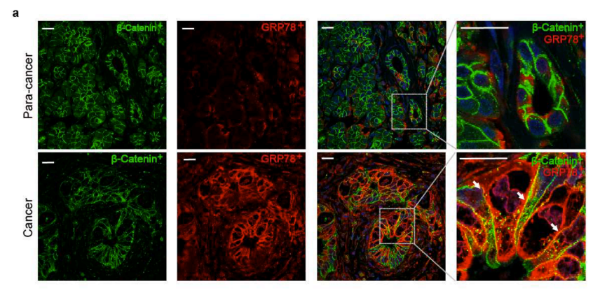
应用: IF
反应种属: Mouse
发表时间: 2023 Oct
-
Citation
-
Isoquercitrin restrains the proliferation and promotes apoptosis of human osteosarcoma cells by inhibiting the Wnt/β-catenin pathway
Author: Wei, Z., Zheng, D., Pi, W., Qiu, Y., Xia, K., & Guo, W.
PMID: 36685044

应用: WB,IHC,IF
反应种属: Human
发表时间: 2023 Jan
-
Citation
-
Fractions of Shen-Sui-Tong-Zhi Formula Enhance Osteogenesis Via Activation of β-Catenin Signaling in Growth Plate Chondrocytes
Author: Xu, R., Zeng, Q., Xia, C., Chen, J., Wang, P., Zhao, S., Yuan, W., Lou, Z., Lin, H., Xia, H., Lv, S., Xu, T., Tong, P., Gu, M., & Jin, H.
PMID: 34630086

应用: WB,IHC
反应种属: Mouse
发表时间: 2021 Sep
-
Citation
-
Moderate DNA hypomethylation suppresses intestinal tumorigenesis by promoting caspase-3 expression and apoptosis. Oncogenesis, 10(5), 38.
Author: Duan, X., Huang, Y., Chen, X., Wang, W., Chen, J., Li, J., Yang, W., Li, J., Wu, Q., & Wong, J.
PMID: 33947834
应用: IHC
反应种属: Mouse
发表时间: 2021 May
-
Citation
-
Klotho Inhibits Proliferation in a RET Fusion Model of Papillary Thyroid Cancer by Regulating the Wnt/β-Catenin Pathway. Cancer management and research, 13, 4791–4802.
Author: Wu, Q., Jiang, L., Wu, J., Dong, H., & Zhao, Y.
PMID: 34168498
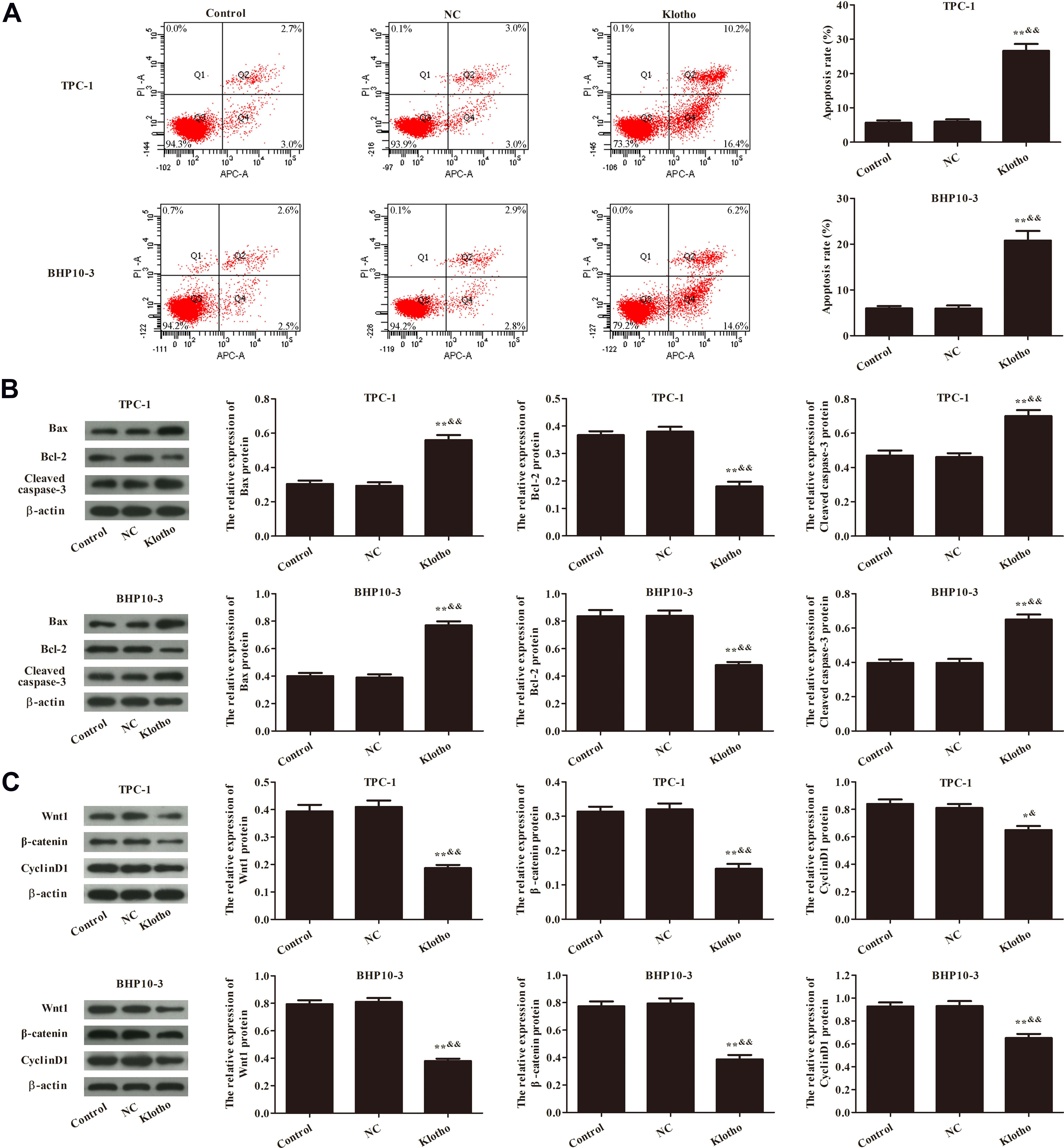
应用: WB
反应种属: Mouse
发表时间: 2021 Jun
-
Citation
Alternative Products
同靶点&同通路的产品
Beta Catenin Rabbit Polyclonal Antibody
Application: WB,IF-Cell,IHC-P
Reactivity: Human,Mouse,Zebrafish,Rat
Conjugate: unconjugated
Phospho-Beta Catenin (T41/S45) Recombinant Rabbit Monoclonal Antibody [JE54-02]
Application: WB,IHC-P
Reactivity: Human,Mouse,Rat
Conjugate: unconjugated
Beta Catenin Mouse Monoclonal Antibody [A6-F8]
Application: WB,IF-Cell,IHC-P,FC,IF-Tissue
Reactivity: Human,Mouse,Rat
Conjugate: unconjugated
Beta Catenin Mouse Monoclonal Antibody [10-C0-B7]
Application: WB,IF-Cell,IHC-P,FC
Reactivity: Human,Mouse,Rat
Conjugate: unconjugated
iFluor™ 488 Conjugated Beta Catenin Recombinant Rabbit Monoclonal Antibody [SA30-04]
Application: IF,ICC
Reactivity: Human,Mouse
Conjugate: iFluor™ 488
Beta Catenin Recombinant Rabbit Monoclonal Antibody [SA30-04]
Application: WB,IHC-P,IF-Tissue,IP,mIHC,IF-Cell,IHC-Fr,FC
Reactivity: Human,Mouse,Rat
Conjugate: unconjugated
Beta Catenin Recombinant Mouse Monoclonal Antibody [A6-F8-R]
Application: WB,IF-Cell,FC
Reactivity: Human,Mouse,Rat
Conjugate: unconjugated
Phospho-Beta Catenin (S33 + S37) Recombinant Rabbit Monoclonal Antibody [JE59-59]
Application: WB
Reactivity: Human,Rat,Mouse
Conjugate: unconjugated


