Ubiquitin Rabbit Polyclonal Antibody


Catalog# ER31212
Ubiquitin Rabbit Polyclonal Antibody
-
WB
-
IF-Cell
-
IHC-P
-
Human
-
Mouse
-
Rat
概述
产品名称
Ubiquitin Rabbit Polyclonal Antibody
抗体类型
Rabbit Polyclonal Antibody
免疫原
Synthetic peptide within human Ubiquitin aa 21-76.
种属反应性
Human, Mouse, Rat
验证应用
WB, IF-Cell, IHC-P
分子量
Predicted band size: 8 kDa
阳性对照
293T cell lysate, HepG2 cell lysate, Hela cell lysate, MCF-7 cell lysate, Hela, HepG2, human tonsil tissue, mouse lung tissue, human kidney tissue.
偶联
unconjugated
RRID
产品特性
形态
Liquid
浓度
1ug/ul
存放说明
Store at +4℃ after thawing. Aliquot store at -20℃ or -80℃. Avoid repeated freeze / thaw cycles.
存储缓冲液
1*PBS (pH7.4), 0.2% BSA, 40% Glycerol. Preservative: 0.05% Sodium Azide.
亚型
IgG
纯化方式
Immunogen affinity purified.
应用稀释度
-
WB
-
1:500-1:1,000
-
IF-Cell
-
1:100-1:200
-
IHC-P
-
1:200-1:1,000
靶点
功能
Ubiquitin is a conserved polypeptide unit that plays an important role in the ubiquitin-proteasome pathway. Ubiquitin exists either covalently attached to another protein, or free (unanchored). When covalently bound, it is conjugated to target proteins via an isopeptide bond either as a monomer (monoubiquitin), a polymer linked via different Lys residues of the ubiquitin (polyubiquitin chains) or a linear polymer linked via the initiator Met of the ubiquitin (linear polyubiquitin chains). Polyubiquitin chains, when attached to a target protein, have different functions depending on the Lys residue of the ubiquitin that is linked: Lys-6-linked may be involved in DNA repair; Lys-11-linked is involved in ERAD (endoplasmic reticulum-associated degradation) and in cell-cycle regulation; Lys-29-linked is involved in lysosomal degradation; Lys-33-linked is involved in kinase modification; Lys-48-linked is involved in protein degradation via the proteasome; Lys-63-linked is involved in endocytosis, DNA-damage responses as well as in signaling processes leading to activation of the transcription factor NF-kappa-B.
背景文献
1. "Lys11-linked ubiquitin chains adopt compact conformations and are preferentially hydrolyzed by the deubiquitinase Cezanne." Bremm A., Freund S.M., Komander D. Nat. Struct. Mol. Biol. 17:939-947(2010)
2. "Polyubiquitin binding and cross-reactivity in the USP domain deubiquitinase USP21." Ye Y., Akutsu M., Reyes-Turcu F., Enchev R.I., Wilkinson K.D., Komander D. EMBO Rep. 12:350-357(2011)
3. "OTU deubiquitinases reveal mechanisms of linkage specificity and enable ubiquitin chain restriction analysis." Mevissen T.E., Hospenthal M.K., Geurink P.P., Elliott P.R., Akutsu M., Arnaudo N., Ekkebus R., Kulathu Y., Wauer T., El Oualid F., Freund S.M., Ovaa H., Komander D.Cell 154:169-184(2013)
序列相似性
Belongs to the ubiquitin family.
翻译后修饰
[Ubiquitin]: Phosphorylated at Ser-65 by PINK1 during mitophagy. Phosphorylated ubiquitin specifically binds and activates parkin (PRKN), triggering mitophagy. Phosphorylation does not affect E1-mediated E2 charging of ubiquitin but affects discharging of E2 enzymes to form polyubiquitin chains. It also affects deubiquitination by deubiquitinase enzymes such as USP30.; [Ubiquitin]: Mono-ADP-ribosylated at the C-terminus by PARP9, a component of the PPAR9-DTX3L complex. ADP-ribosylation requires processing by E1 and E2 enzymes and prevents ubiquitin conjugation to substrates such as histones.
亚细胞定位
Cytoplasm, nucleus, Membrane, Mitochondrion, Mitochondrion outer membrane.
UNIPROT #
别名
Epididymis secretory protein Li 50 antibody
FLJ25987 antibody
HEL S 50 antibody
MGC8385 antibody
Polyubiquitin B antibody
RPS 27A antibody
RPS27A antibody
UBA 52 antibody
UBA 80 antibody
UBA52 antibody
展开图片
-
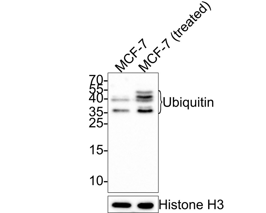
Western blot analysis of Ubiquitin on different lysates with Rabbit anti-Ubiquitin antibody (ER31212) at 1/500 dilution.
Lane 1: MCF-7 cell lysate
Lane 2: MCF-7 cell lysate treated with 50 µM MG132 for 1.5h
Lysates/proteins at 10 µg/Lane.
Predicted band size: 8 kDa
Exposure time: 2 minutes;
15% SDS-PAGE gel.
Proteins were transferred to a PVDF membrane and blocked with 5% NFDM/TBST for 1 hour at room temperature. The primary antibody (ER31212) at 1/500 dilution was used in 5% NFDM/TBST at room temperature for 2 hours. Goat Anti-Rabbit IgG - HRP Secondary Antibody (HA1001) at 1:300,000 dilution was used for 1 hour at room temperature. -
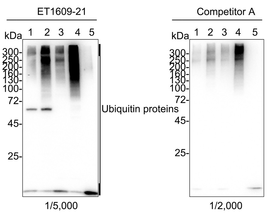
Western blot analysis of Ubiquitin on different cell lysates using anti-Ubiquitin antibody at 1/1,000 dilution.
Positive control:
Lane 1: 293T cell lysate
Lane 2: HepG2 cell lysate
Lane 3: Hela cell lysate
Lane 4: MCF-7 cell lysate -
ICC staining Ubiquitin in Hela cells (green). Cells were fixed in paraformaldehyde, permeabilised with 0.25% Triton X100/PBS.
-
ICC staining Ubiquitin in HepG2 cells (green). Cells were fixed in paraformaldehyde, permeabilised with 0.25% Triton X100/PBS.
-
ICC staining Ubiquitin in MCF-7 cells (green). Cells were fixed in paraformaldehyde, permeabilised with 0.25% Triton X100/PBS.
-
Immunohistochemical analysis of paraffin-embedded mouse lung tissue with Rabbit anti-Ubiquitin antibody (ER31212) at 1/1,000 dilution.
The section was pre-treated using heat mediated antigen retrieval with sodium citrate buffer (pH 6.0) for 2 minutes. The tissues were blocked in 1% BSA for 20 minutes at room temperature, washed with ddH2O and PBS, and then probed with the primary antibody (ER31212) at 1/1,000 dilution for 1 hour at room temperature. The detection was performed using an HRP conjugated compact polymer system. DAB was used as the chromogen. Tissues were counterstained with hematoxylin and mounted with DPX. -
Immunohistochemical analysis of paraffin-embedded human kidney tissue with Rabbit anti-Ubiquitin antibody (ER31212) at 1/1,000 dilution.
The section was pre-treated using heat mediated antigen retrieval with sodium citrate buffer (pH 6.0) for 2 minutes. The tissues were blocked in 1% BSA for 20 minutes at room temperature, washed with ddH2O and PBS, and then probed with the primary antibody (ER31212) at 1/1,000 dilution for 1 hour at room temperature. The detection was performed using an HRP conjugated compact polymer system. DAB was used as the chromogen. Tissues were counterstained with hematoxylin and mounted with DPX. -
Immunocytochemistry analysis of NIH/3T3 cells labeling Ubiquitin with Rabbit anti-Ubiquitin antibody (ER31212) at 1/100 dilution.
Cells were fixed in 4% paraformaldehyde for 20 minutes at room temperature, permeabilized with 0.1% Triton X-100 in PBS for 5 minutes at room temperature, then blocked with 1% BSA in 10% negative goat serum for 1 hour at room temperature. Cells were then incubated with Rabbit anti-Ubiquitin antibody (ER31212) at 1/100 dilution in 1% BSA in PBST overnight at 4 ℃. Goat Anti-Rabbit IgG H&L (iFluor™ 488, HA1121) was used as the secondary antibody at 1/1,000 dilution. PBS instead of the primary antibody was used as the secondary antibody only control. Nuclear DNA was labelled in blue with DAPI.
Beta tubulin (M1305-2, red) was stained at 1/100 dilution overnight at +4℃. Goat Anti-Mouse IgG H&L (iFluor™ 594, HA1126) was used as the secondary antibody at 1/1,000 dilution. -
Immunocytochemistry analysis of PC-12 cells labeling Ubiquitin with Rabbit anti-Ubiquitin antibody (ER31212) at 1/100 dilution.
Cells were fixed in 4% paraformaldehyde for 20 minutes at room temperature, permeabilized with 0.1% Triton X-100 in PBS for 5 minutes at room temperature, then blocked with 1% BSA in 10% negative goat serum for 1 hour at room temperature. Cells were then incubated with Rabbit anti-Ubiquitin antibody (ER31212) at 1/100 dilution in 1% BSA in PBST overnight at 4 ℃. Goat Anti-Rabbit IgG H&L (iFluor™ 488, HA1121) was used as the secondary antibody at 1/1,000 dilution. PBS instead of the primary antibody was used as the secondary antibody only control. Nuclear DNA was labelled in blue with DAPI.
Beta tubulin (M1305-2, red) was stained at 1/100 dilution overnight at +4℃. Goat Anti-Mouse IgG H&L (iFluor™ 594, HA1126) was used as the secondary antibody at 1/1,000 dilution.
Please note: All products are "FOR RESEARCH USE ONLY AND ARE NOT INTENDED FOR DIAGNOSTIC OR THERAPEUTIC USE"
同靶点&同通路的产品

Ubiquitin Mouse Monoclonal Antibody [6G6]
Application: WB,IHC-P
Reactivity: Human,Rat,Mouse
Conjugate: unconjugated