STAT6 Recombinant Rabbit Monoclonal Antibody [SY02-72]


Catalog# ET1607-61
STAT6 Recombinant Rabbit Monoclonal Antibody [SY02-72]
-
WB
-
IF-Cell
-
IF-Tissue
-
IHC-P
-
IP
-
FC
-
Human
-
Mouse
-
Rat
-
unconjugated
概述
产品名称
STAT6 Recombinant Rabbit Monoclonal Antibody [SY02-72]
抗体类型
Recombinant Rabbit monoclonal Antibody
免疫原
Synthetic peptide within Human STAT6 aa 800 to the C-terminus.
种属反应性
Human, Mouse, Rat
验证应用
WB, IF-Cell, IF-Tissue, IHC-P, IP, FC
分子量
Predicted band size: 94 kDa
阳性对照
Raji cell lysate, RAW264.7 cell lysate, mouse kidney tissue, rat kidney tissue, human kidney tissue, mouse lung tissue, mouse stomach tissue, RAW264.7, Hela, AGS, NIH/3T3.
偶联
unconjugated
克隆号
SY02-72
RRID
产品特性
形态
Liquid
浓度
存放说明
Shipped at 4℃. Store at +4℃ short term (1-2 weeks). It is recommended to aliquot into single-use upon delivery. Store at -20℃ long term.
存储缓冲液
1*TBS (pH7.4), 0.05% BSA, 40% Glycerol. Preservative: 0.05% Sodium Azide.
亚型
IgG
纯化方式
Protein A affinity purified.
应用稀释度
-
WB
-
1:2,000
-
IF-Cell
-
1:50
-
IF-Tissue
-
1:50
-
IHC-P
-
1:50-1:200
-
IP
-
Use at an assay dependent concentration.
-
FC
-
1:1,000
靶点
功能
Signal transducer and activator of transcription 6 (STAT6) is a transcription factor that belongs to the Signal Transducer and Activator of Transcription (STAT) family of proteins. The proteins of STAT family transmit signals from a receptor complex to the nucleus and activate gene expression. Similarly as other STAT family proteins, STAT6 is also activated by growth factors and cytokines. STAT6 is mainly activated by cytokines interleukin-4 and interleukin-13. STAT6-mediated signaling pathway is required for the development of T-helper type 2 (Th2) cells and Th2 immune response. Activation of STAT6 signaling pathway is necessary in macrophage function, and is required for the M2 subtype activation of macrophages. STAT6 is also involved in IL4 signaling in B cells, and STAT6 determines the levels of CD20 on the surface of normal and malignant B lymphocytes. STAT6 also plays a critical role in Th2 lung inflammatory responses including clearance of parasitic infections and in the pathogenesis of asthma. Th2-cell derived cytokines as IL-4 and IL-13 induce the production of IgE which is a major mediator in allergic response.
背景文献
1. Zheng, C. et al. 2015. CD11b regulates obesity-induced insulin resistance via limiting alternative activation and proliferation of adipose tissue macrophages. Proc. Natl. Acad. Sci. U.S.A.. 112: E7239-48.
2. Carlson, TJ. et al. 2014. Halofuginone-induced amino acid starvation regulates Stat3-dependent Th17 effector function and reduces established autoimmune inflammation. J. Immunol.. 192: 2167-76.
序列相似性
Belongs to the transcription factor STAT family.
翻译后修饰
Tyrosine phosphorylated on Tyr-641 following stimulation by IL4/interleukin-4. Tyrosine phosphorylated following stimulation by IL3/interleukin-3 (By similarity). Dephosphorylation on tyrosine residues by PTPN2 negatively regulates the IL4/interleukin-4 mediated signaling.; Mono-ADP-ribosylated by PARP14.
亚细胞定位
Cytoplasm, Nucleus.
别名
12S1644 antibody
D12S1644 antibody
IL 4 STAT antibody
IL-4 Stat antibody
IL4 STAT antibody
Interleukin 4 Induced antibody
Interleukin 4 Induced Transcription Factor IL4 STAT antibody
Signal transducer and activator of transcription 6 antibody
Signal Transducer And Activator Of Transcription 6 Interleukin 4 Induced antibody
Signal Transducer And Activator Of Transcription 6 Nirs Variant 1 antibody
展开图片
-
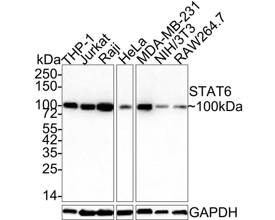
Western blot analysis of STAT6 on different lysates with Rabbit anti-STAT6 antibody (ET1607-61) at 1/2,000 dilution.
Lane 1: Raji cell lysate
Lane 2: RAW264.7 cell lysate
Lysates/proteins at 30 µg/Lane.
Predicted band size: 94 kDa
Observed band size: 94 kDa
Exposure time: 3 minutes; ECL: K1801;
4-20% SDS-PAGE gel.
Proteins were transferred to a PVDF membrane and blocked with 5% NFDM/TBST for 1 hour at room temperature. The primary antibody (ET1607-61) at 1/2,000 dilution was used in 5% NFDM/TBST at 4℃ overnight. Goat Anti-Rabbit IgG - HRP Secondary Antibody (HA1001) at 1/50,000 dilution was used for 1 hour at room temperature. -
☑ Knockdown (KD)
Western blot analysis of STAT6 on different lysates with Rabbit anti-STAT6 antibody (ET1607-61) at 1/500 dilution.
Lane 1: Hela-si NT cell lysate
Lane 2: Hela-si STAT6 cell lysate
Lysates/proteins at 10 µg/Lane.
Predicted band size: 94 kDa
Observed band size: 94 kDa
Exposure time: 3 minutes;
4-20% SDS-PAGE gel.
ET1607-61 was shown to specifically react with STAT6 in Hela-si NT cells. Weakened band was observed when Hela-si STAT6 sample was tested. Hela-si NT and Hela-si STAT6 samples were subjected to SDS-PAGE. Proteins were transferred to a PVDF membrane and blocked with 5% NFDM in TBST for 1 hour at room temperature. The primary antibody (ET1607-61, 1/500) and Loading control antibody (Rabbit anti-GAPDH, ET1601-4, 1/10,000) were used in 5% BSA at room temperature for 2 hours. Goat Anti-rabbit IgG-HRP Secondary Antibody (HA1001) at 1:200,000 dilution was used for 1 hour at room temperature. -
Immunohistochemical analysis of paraffin-embedded mouse kidney tissue with Rabbit anti-STAT6 antibody (ET1607-61) at 1/200 dilution.
The section was pre-treated using heat mediated antigen retrieval with Tris-EDTA buffer (pH 9.0) for 20 minutes. The tissues were blocked in 1% BSA for 20 minutes at room temperature, washed with ddH2O and PBS, and then probed with the primary antibody (ET1607-61) at 1/200 dilution for 1 hour at room temperature. The detection was performed using an HRP conjugated compact polymer system. DAB was used as the chromogen. Tissues were counterstained with hematoxylin and mounted with DPX. -
Immunohistochemical analysis of paraffin-embedded rat kidney tissue with Rabbit anti-STAT6 antibody (ET1607-61) at 1/200 dilution.
The section was pre-treated using heat mediated antigen retrieval with Tris-EDTA buffer (pH 9.0) for 20 minutes. The tissues were blocked in 1% BSA for 20 minutes at room temperature, washed with ddH2O and PBS, and then probed with the primary antibody (ET1607-61) at 1/200 dilution for 1 hour at room temperature. The detection was performed using an HRP conjugated compact polymer system. DAB was used as the chromogen. Tissues were counterstained with hematoxylin and mounted with DPX. -
Immunohistochemical analysis of paraffin-embedded human kidney tissue with Rabbit anti-STAT6 antibody (ET1607-61) at 1/50 dilution.
The section was pre-treated using heat mediated antigen retrieval with Tris-EDTA buffer (pH 9.0) for 20 minutes. The tissues were blocked in 1% BSA for 20 minutes at room temperature, washed with ddH2O and PBS, and then probed with the primary antibody (ET1607-61) at 1/50 dilution for 1 hour at room temperature. The detection was performed using an HRP conjugated compact polymer system. DAB was used as the chromogen. Tissues were counterstained with hematoxylin and mounted with DPX. -
Immunohistochemical analysis of paraffin-embedded mouse lung tissue with Rabbit anti-STAT6 antibody (ET1607-61) at 1/20 dilution.
The section was pre-treated using heat mediated antigen retrieval with Tris-EDTA buffer (pH 9.0) for 20 minutes. The tissues were blocked in 1% BSA for 20 minutes at room temperature, washed with ddH2O and PBS, and then probed with the primary antibody (ET1607-61) at 1/20 dilution for 1 hour at room temperature. The detection was performed using an HRP conjugated compact polymer system. DAB was used as the chromogen. Tissues were counterstained with hematoxylin and mounted with DPX. -
Immunohistochemical analysis of paraffin-embedded mouse stomach tissue with Rabbit anti-STAT6 antibody (ET1607-61) at 1/50 dilution.
The section was pre-treated using heat mediated antigen retrieval with Tris-EDTA buffer (pH 9.0) for 20 minutes. The tissues were blocked in 1% BSA for 20 minutes at room temperature, washed with ddH2O and PBS, and then probed with the primary antibody (ET1607-61) at 1/50 dilution for 1 hour at room temperature. The detection was performed using an HRP conjugated compact polymer system. DAB was used as the chromogen. Tissues were counterstained with hematoxylin and mounted with DPX. -
Flow cytometric analysis of RAW264.7 cells labeling STAT6.
Cells were fixed and permeabilized. Then stained with the primary antibody (ET1607-61, 1/1,000) (red) compared with Rabbit IgG Isotype Control (green). After incubation of the primary antibody at +4℃ for an hour, the cells were stained with a iFluor™ 488 conjugate-Goat anti-Rabbit IgG Secondary antibody (HA1121) at 1/1,000 dilution for 30 minutes at +4℃. Unlabelled sample was used as a control (cells without incubation with primary antibody; black). -
Immunocytochemistry analysis of Hela cells labeling STAT6 with Rabbit anti-STAT6 antibody (ET1607-61) at 1/50 dilution.
Cells were fixed in 4% paraformaldehyde for 10 minutes at 37 ℃, permeabilized with 0.05% Triton X-100 in PBS for 20 minutes, and then blocked with 2% negative goat serum for 30 minutes at room temperature. Cells were then incubated with Rabbit anti-STAT6 antibody (ET1607-61) at 1/50 dilution in 2% negative goat serum overnight at 4 ℃. Goat Anti-Rabbit IgG H&L (iFluor™ 488, HA1121) was used as the secondary antibody at 1/1,000 dilution. PBS instead of the primary antibody was used as the secondary antibody only control. Nuclear DNA was labelled in blue with DAPI. -
Immunocytochemistry analysis of AGS cells labeling STAT6 with Rabbit anti-STAT6 antibody (ET1607-61) at 1/50 dilution.
Cells were fixed in 4% paraformaldehyde for 10 minutes at 37 ℃, permeabilized with 0.05% Triton X-100 in PBS for 20 minutes, and then blocked with 2% negative goat serum for 30 minutes at room temperature. Cells were then incubated with Rabbit anti-STAT6 antibody (ET1607-61) at 1/50 dilution in 2% negative goat serum overnight at 4 ℃. Goat Anti-Rabbit IgG H&L (iFluor™ 488, HA1121) was used as the secondary antibody at 1/1,000 dilution. PBS instead of the primary antibody was used as the secondary antibody only control. Nuclear DNA was labelled in blue with DAPI. -
Immunocytochemistry analysis of NIH/3T3 cells labeling STAT6 with Rabbit anti-STAT6 antibody (ET1607-61) at 1/50 dilution.
Cells were fixed in 4% paraformaldehyde for 10 minutes at 37 ℃, permeabilized with 0.05% Triton X-100 in PBS for 20 minutes, and then blocked with 2% negative goat serum for 30 minutes at room temperature. Cells were then incubated with Rabbit anti-STAT6 antibody (ET1607-61) at 1/50 dilution in 2% negative goat serum overnight at 4 ℃. Goat Anti-Rabbit IgG H&L (iFluor™ 488, HA1121) was used as the secondary antibody at 1/1,000 dilution. PBS instead of the primary antibody was used as the secondary antibody only control. Nuclear DNA was labelled in blue with DAPI.
请注意: All products are "FOR RESEARCH USE ONLY AND ARE NOT INTENDED FOR DIAGNOSTIC OR THERAPEUTIC USE"
同靶点 & 同通路的产品
STAT6 Recombinant Rabbit Monoclonal Antibody [SY13-09]
Application: WB,IF-Cell,IHC-P,IP,FC,IF-Tissue
Reactivity: Human,Mouse,Rat
Conjugate: unconjugated

 浙公网安备 33019202000643号