PAX8 Recombinant Rabbit Monoclonal Antibody [JE80-01]


Catalog# HA720112
PAX8 Recombinant Rabbit Monoclonal Antibody [JE80-01]
-
WB
-
IHC-P
-
IF-Cell
-
Human
-
Mouse
概述
产品名称
PAX8 Recombinant Rabbit Monoclonal Antibody [JE80-01]
抗体类型
Recombinant Rabbit monoclonal Antibody
免疫原
Recombinant protein within Human PAX8 aa 120-450.
种属反应性
Human, Mouse
验证应用
WB, IHC-P, IF-Cell
分子量
Predicted band size: 48 kDa
阳性对照
NIH: OVCAR-3 cell lysates, SKOV-3 cell lysates, SKOV-3, human thyroid tissue, human kidney tissue, human thyroid carcinoma tissue, human ovary carcinoma tissue, human renal clear cell carcinoma tissue, mouse thyroid tissue, mouse kidney tissue, rat kidney tissue.
偶联
unconjugated
克隆号
JE80-01
RRID
产品特性
形态
Liquid
浓度
1ug/ul
存放说明
Store at +4℃ after thawing. Aliquot store at -20℃. Avoid repeated freeze / thaw cycles.
存储缓冲液
1*TBS (pH7.4), 0.05% BSA, 40% Glycerol. Preservative: 0.05% Sodium Azide.
亚型
IgG
纯化方式
Protein A affinity purified.
应用稀释度
-
WB
-
1:1,000
-
IHC-P
-
1:500-1:5,000
-
IF-Cell
-
1:100
靶点
功能
PAX8 is a transcription factor crucial to the organogenesis and development of the thyroid gland, urogenital tract, placenta and inner ear. In the thyroid, PAX8 is a master gene that regulates maintenance of the differentiated thyroid follicular cell phenotype, where it controls and activates the transcription of the main proteins responsible for the functional activity of follicular cells such as thyroglobulin, thyroperoxidase and sodium/iodide symporter. In the developing kidney PAX8 is important for renal vescicle formation. PAX8 regulates the expression of the WT1 gene. PAX8 appears to be currently the most sensitive and specific marker for renal cell carcinoma and ovarian non-mucinous carcinoma. Nuclear transcriptional regulator in the paired-box family expressed during organogenesis of the thyroid gland, kidney, and Müllerian tract. Chromosomal localization 2q13, Molecular weight of the unprocessed precursor 48 kDa, of five isoforms 31-42 kDa.
背景文献
1. Wang M et al. PAX2 and PAX8 reliably distinguishes ovarian serous tumors from mucinous tumors. Appl Immunohistochem Mol Morphol 23:280-7 (2015).
2. Sicking EM et al. Subtotal ablation of parietal epithelial cells induces crescent formation. J Am Soc Nephrol 23:629-40 (2012).
亚细胞定位
Nucleus.
UNIPROT #
别名
OTTHUMP00000158659 antibody
OTTHUMP00000158660 antibody
OTTHUMP00000203723 antibody
OTTHUMP00000203724 antibody
Paired box 8 antibody
Paired box gene 8 antibody
paired box homeotic gene 8 antibody
Paired box protein Pax 8 antibody
Paired box protein Pax-8 antibody
Paired domain gene 8 antibody
展开图片
-
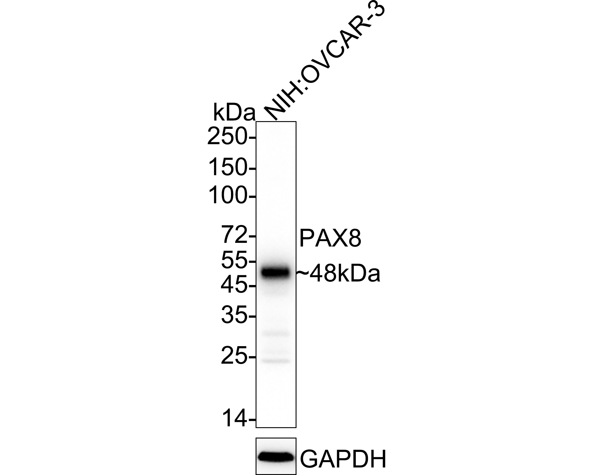
Western blot analysis of PAX8 on NIH: OVCAR-3 cell lysates with Rabbit anti-PAX8 antibody (HA720112) at 1/1,000 dilution.
Lysates/proteins at 20 µg/Lane.
Predicted band size: 48 kDa
Observed band size: 48 kDa
Exposure time: 18 seconds; ECL: K1801;
4-20% SDS-PAGE gel.
Proteins were transferred to a PVDF membrane and blocked with 5% NFDM/TBST for 1 hour at room temperature. The primary antibody (HA720112) at 1/1,000 dilution was used in 5% NFDM/TBST at 4℃ overnight. Goat Anti-Rabbit IgG - HRP Secondary Antibody (HA1001) at 1/50,000 dilution was used for 1 hour at room temperature. -
Western blot analysis of PAX8 on SKOV-3 cell lysates with Rabbit anti-PAX8 antibody (HA720112) at 1/1,000 dilution.
Lysates/proteins at 10 µg/Lane.
Predicted band size: 48 kDa
Observed band size: 48 kDa
Exposure time: 2 minutes;
8% SDS-PAGE gel.
Proteins were transferred to a PVDF membrane and blocked with 5% NFDM/TBST for 1 hour at room temperature. The primary antibody (HA720112) at 1/1,000 dilution was used in 5% NFDM/TBST at room temperature for 2 hours. Goat Anti-Rabbit IgG - HRP Secondary Antibody (HA1001) at 1:300,000 dilution was used for 1 hour at room temperature. -
Immunocytochemistry analysis of SKOV-3 cells labeling PAX8 with Rabbit anti-PAX8 antibody (HA720112) at 1/100 dilution.
Cells were fixed in 4% paraformaldehyde for 10 minutes at 37 ℃, permeabilized with 0.05% Triton X-100 in PBS for 20 minutes, and then blocked with 2% negative goat serum for 30 minutes at room temperature. Cells were then incubated with Rabbit anti-PAX8 antibody (HA720112) at 1/100 dilution in 2% negative goat serum overnight at 4 ℃. Goat Anti-Rabbit IgG H&L (iFluor™ 488, HA1121) was used as the secondary antibody at 1/1,000 dilution. PBS instead of the primary antibody was used as the secondary antibody only control. Nuclear DNA was labelled in blue with DAPI. -
Immunohistochemical analysis of paraffin-embedded human thyroid tissue with Rabbit anti-PAX8 antibody (HA720112) at 1/5,000 dilution.
The section was pre-treated using heat mediated antigen retrieval with sodium citrate buffer (pH 6.0) for 2 minutes. The tissues were blocked in 1% BSA for 20 minutes at room temperature, washed with ddH2O and PBS, and then probed with the primary antibody (HA720112) at 1/5,000 dilution for 1 hour at room temperature. The detection was performed using an HRP conjugated compact polymer system. DAB was used as the chromogen. Tissues were counterstained with hematoxylin and mounted with DPX. -
Immunohistochemical analysis of paraffin-embedded human kidney tissue with Rabbit anti-PAX8 antibody (HA720112) at 1/2,000 dilution.
The section was pre-treated using heat mediated antigen retrieval with sodium citrate buffer (pH 6.0) for 2 minutes. The tissues were blocked in 1% BSA for 20 minutes at room temperature, washed with ddH2O and PBS, and then probed with the primary antibody (HA720112) at 1/2,000 dilution for 1 hour at room temperature. The detection was performed using an HRP conjugated compact polymer system. DAB was used as the chromogen. Tissues were counterstained with hematoxylin and mounted with DPX. -
Immunohistochemical analysis of paraffin-embedded human thyroid carcinoma tissue with Rabbit anti-PAX8 antibody (HA720112) at 1/5,000 dilution.
The section was pre-treated using heat mediated antigen retrieval with sodium citrate buffer (pH 6.0) for 2 minutes. The tissues were blocked in 1% BSA for 20 minutes at room temperature, washed with ddH2O and PBS, and then probed with the primary antibody (HA720112) at 1/5,000 dilution for 1 hour at room temperature. The detection was performed using an HRP conjugated compact polymer system. DAB was used as the chromogen. Tissues were counterstained with hematoxylin and mounted with DPX. -
Immunohistochemical analysis of paraffin-embedded human ovary carcinoma tissue with Rabbit anti-PAX8 antibody (HA720112) at 1/1,000 dilution.
The section was pre-treated using heat mediated antigen retrieval with sodium citrate buffer (pH 6.0) for 2 minutes. The tissues were blocked in 1% BSA for 20 minutes at room temperature, washed with ddH2O and PBS, and then probed with the primary antibody (HA720112) at 1/1,000 dilution for 1 hour at room temperature. The detection was performed using an HRP conjugated compact polymer system. DAB was used as the chromogen. Tissues were counterstained with hematoxylin and mounted with DPX. -
Immunohistochemical analysis of paraffin-embedded human renal clear cell carcinoma tissue with Rabbit anti-PAX8 antibody (HA720112) at 1/500 dilution.
The section was pre-treated using heat mediated antigen retrieval with sodium citrate buffer (pH 6.0) for 2 minutes. The tissues were blocked in 1% BSA for 20 minutes at room temperature, washed with ddH2O and PBS, and then probed with the primary antibody (HA720112) at 1/500 dilution for 1 hour at room temperature. The detection was performed using an HRP conjugated compact polymer system. DAB was used as the chromogen. Tissues were counterstained with hematoxylin and mounted with DPX. -
Immunohistochemical analysis of paraffin-embedded mouse thyroid tissue with Rabbit anti-PAX8 antibody (HA720112) at 1/2,000 dilution.
The section was pre-treated using heat mediated antigen retrieval with sodium citrate buffer (pH 6.0) for 2 minutes. The tissues were blocked in 1% BSA for 20 minutes at room temperature, washed with ddH2O and PBS, and then probed with the primary antibody (HA720112) at 1/2,000 dilution for 1 hour at room temperature. The detection was performed using an HRP conjugated compact polymer system. DAB was used as the chromogen. Tissues were counterstained with hematoxylin and mounted with DPX. -
Immunohistochemical analysis of paraffin-embedded mouse kidney tissue with Rabbit anti-PAX8 antibody (HA720112) at 1/500 dilution.
The section was pre-treated using heat mediated antigen retrieval with sodium citrate buffer (pH 6.0) for 2 minutes. The tissues were blocked in 1% BSA for 20 minutes at room temperature, washed with ddH2O and PBS, and then probed with the primary antibody (HA720112) at 1/500 dilution for 1 hour at room temperature. The detection was performed using an HRP conjugated compact polymer system. DAB was used as the chromogen. Tissues were counterstained with hematoxylin and mounted with DPX. -
Immunohistochemical analysis of paraffin-embedded rat kidney tissue with Rabbit anti-PAX8 antibody (HA720112) at 1/500 dilution.
The section was pre-treated using heat mediated antigen retrieval with sodium citrate buffer (pH 6.0) for 2 minutes. The tissues were blocked in 1% BSA for 20 minutes at room temperature, washed with ddH2O and PBS, and then probed with the primary antibody (HA720112) at 1/500 dilution for 1 hour at room temperature. The detection was performed using an HRP conjugated compact polymer system. DAB was used as the chromogen. Tissues were counterstained with hematoxylin and mounted with DPX.
Please note: All products are "FOR RESEARCH USE ONLY AND ARE NOT INTENDED FOR DIAGNOSTIC OR THERAPEUTIC USE"