Lactate Dehydrogenase Rabbit Polyclonal Antibody


Catalog# ER00702
Lactate Dehydrogenase Rabbit Polyclonal Antibody
-
WB
-
IHC-P
-
FC
-
Human
-
Mouse
-
Rat
概述
产品名称
Lactate Dehydrogenase Rabbit Polyclonal Antibody
抗体类型
Rabbit Polyclonal Antibody
免疫原
Synthetic peptide within Human LDHA aa1-50 / 332.
种属反应性
Human, Mouse, Rat
验证应用
WB, IHC-P, FC
分子量
Predicted band size: 37 kDa
阳性对照
A431 cell lysate, MCF7 cell lysate, NIH/3T3 cell lysate, RAW264.7 cell lysate, C6 cell lysate, PC-12 cell lysate, Wild-type Hela whole cell lysate, rat liver tissue, rat skeletal muscle tissue, human liver tissue, human breast carcinoma tissue, mouse liver tissue.
偶联
unconjugated
RRID
产品特性
形态
Liquid
浓度
1ug/ul
存放说明
Store at +4℃ after thawing. Aliquot store at -20℃ or -80℃. Avoid repeated freeze / thaw cycles.
存储缓冲液
1*PBS (pH7.4), 0.2% BSA, 40% Glycerol. Preservative: 0.05% Sodium Azide.
亚型
IgG
纯化方式
Immunogen affinity purified.
应用稀释度
-
WB
-
1:5,000
-
IHC-P
-
1:200
-
FC
-
1:100-1:200
发表文章中的应用
靶点
功能
Lactate dehydrogenase (LDH) is an enzyme present in a wide variety of organisms, including plants and animals. It catalyses the interconversion of pyruvate and lactate with concomitant interconversion of NADH and NAD+. In medicine, LDH is often used as a marker of tissue breakdown as LDH is abundant in red blood cells and can function as a marker for hemolysis. In mammals, three types of LDH subunits (35 kDa) are encoded by the genes Ldh-A, Ldh-B, and Ldh-C. Lactate dehydrogenase B (LDH-B, heart subunit, LDH-H) is involved in the conversion of L-lactate and NAD to pryruvate and NADH and it is predominantly localized in the heart tissue. Similar to other LDH subunit, LDH-B is considered to be an important marker for germ cell tumor.
背景文献
1. Miskimins WK et al. Synergistic anti-cancer effect of phenformin and oxamate. PLoS One 9:e85576 (2014)
2. Peng X et al. Autophagy promotes paclitaxel resistance of cervical cancer cells: involvement of Warburg effect activated hypoxia-induced factor 1-a-mediated signaling. Cell Death Dis 5:e1367 (2014)
序列相似性
Belongs to the LDH/MDH superfamily. LDH family.
翻译后修饰
ISGylated.
亚细胞定位
Cytoplasm.
UNIPROT #
别名
Cell proliferation-inducing gene 19 protein antibody
GSD11 antibody
L lactate dehydrogenase A chain antibody
L-lactate dehydrogenase A chain antibody
l7R2 antibody
Lactate dehydrogenase 1, A chain antibody
Lactate dehydrogenase A antibody
Lactate dehydrogenase A4 antibody
Lactate dehydrogenase M antibody
LDH A antibody
展开图片
-
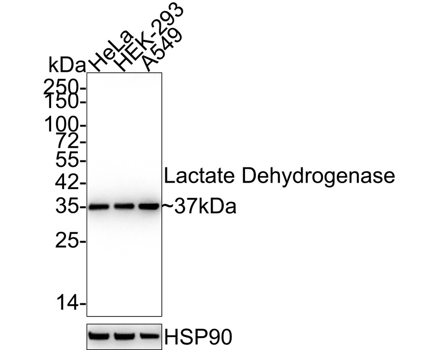
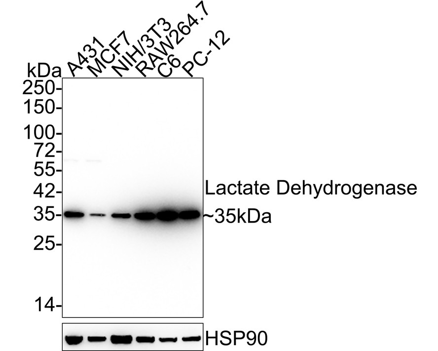
Western blot analysis of Lactate Dehydrogenase on different lysates with Rabbit anti-Lactate Dehydrogenase antibody (ER00702) at 1/5,000 dilution.
Lane 1: A431 cell lysate
Lane 2: MCF7 cell lysate
Lane 3: NIH/3T3 cell lysate
Lane 4: RAW264.7 cell lysate
Lane 5: C6 cell lysate
Lane 6: PC-12 cell lysate
Lysates/proteins at 15 µg/Lane.
Predicted band size: 37 kDa
Observed band size: 35 kDa
Exposure time: 20 seconds;
4-20% SDS-PAGE gel.
Proteins were transferred to a PVDF membrane and blocked with 5% NFDM/TBST for 1 hour at room temperature. The primary antibody (ER00702) at 1/5,000 dilution was used in 5% NFDM/TBST at 4℃ overnight. Goat Anti-Rabbit IgG - HRP Secondary Antibody (HA1001) at 1/50,000 dilution was used for 1 hour at room temperature. -
All lanes: Western blot analysis of LDHA with anti-LDHA antibody (ER00702) at 1:500 dilution.
Lane 1: Wild-type Hela whole cell lysate (10 µg).
Lane 2/3: LDHA knockdown Hela whole cell lysate (10 µg).
ER00702 was shown to specifically react with LDHA in wild-type Hela cells. Weakened bands were observed when LDHA knockdown samples were tested. Wild-type and LDHA knockdown samples were subjected to SDS-PAGE. Proteins were transferred to a PVDF membrane and blocked with 5% NFDM in TBST for 1 hour at room temperature. The primary antibody (ER00702, 1:500) was used in 5% BSA at room temperature for 2 hours. Goat Anti-Rabbit IgG-HRP Secondary Antibody (HA1001) at 1:300,000 dilution was used for 1 hour at room temperature. -
Immunohistochemical analysis of paraffin-embedded rat liver tissue using anti-Lactate Dehydrogenase antibody. The section was pre-treated using heat mediated antigen retrieval with Tris-EDTA buffer (pH 9.0) for 20 minutes.The tissues were blocked in 1% BSA for 30 minutes at room temperature, washed with ddH2O and PBS, and then probed with the primary antibody (ER00702, 1/50) for 30 minutes at room temperature. The detection was performed using an HRP conjugated compact polymer system. DAB was used as the chromogen. Tissues were counterstained with hematoxylin and mounted with DPX.
-
Immunohistochemical analysis of paraffin-embedded rat skeletal muscle tissue using anti-Lactate Dehydrogenase antibody. The section was pre-treated using heat mediated antigen retrieval with Tris-EDTA buffer (pH 9.0) for 20 minutes.The tissues were blocked in 1% BSA for 30 minutes at room temperature, washed with ddH2O and PBS, and then probed with the primary antibody (ER00702, 1/50) for 30 minutes at room temperature. The detection was performed using an HRP conjugated compact polymer system. DAB was used as the chromogen. Tissues were counterstained with hematoxylin and mounted with DPX.
-
Immunohistochemical analysis of paraffin-embedded human liver tissue using anti-Lactate Dehydrogenase antibody. The section was pre-treated using heat mediated antigen retrieval with Tris-EDTA buffer (pH 9.0) for 20 minutes.The tissues were blocked in 1% BSA for 30 minutes at room temperature, washed with ddH2O and PBS, and then probed with the primary antibody (ER00702, 1/50) for 30 minutes at room temperature. The detection was performed using an HRP conjugated compact polymer system. DAB was used as the chromogen. Tissues were counterstained with hematoxylin and mounted with DPX.
-
Immunohistochemical analysis of paraffin-embedded human breast carcinoma tissue using anti-Lactate Dehydrogenase antibody. The section was pre-treated using heat mediated antigen retrieval with Tris-EDTA buffer (pH 9.0) for 20 minutes.The tissues were blocked in 1% BSA for 30 minutes at room temperature, washed with ddH2O and PBS, and then probed with the primary antibody (ER00702, 1/50) for 30 minutes at room temperature. The detection was performed using an HRP conjugated compact polymer system. DAB was used as the chromogen. Tissues were counterstained with hematoxylin and mounted with DPX.
-
Immunohistochemical analysis of paraffin-embedded mouse liver tissue using anti-Lactate Dehydrogenase antibody. The section was pre-treated using heat mediated antigen retrieval with Tris-EDTA buffer (pH 9.0) for 20 minutes.The tissues were blocked in 1% BSA for 30 minutes at room temperature, washed with ddH2O and PBS, and then probed with the primary antibody (ER00702, 1/50) for 30 minutes at room temperature. The detection was performed using an HRP conjugated compact polymer system. DAB was used as the chromogen. Tissues were counterstained with hematoxylin and mounted with DPX.
Please note: All products are "FOR RESEARCH USE ONLY AND ARE NOT INTENDED FOR DIAGNOSTIC OR THERAPEUTIC USE"

Alternative Products

Lactate Dehydrogenase A Mouse Monoclonal Antibody [20-179]
Application: IHC-P
Reactivity: Human,Mouse
Conjugate: unconjugated
同靶点&同通路的产品