LAMP2a Recombinant Rabbit Monoclonal Antibody [SA46-01]


Catalog# ET1601-24
LAMP2a Recombinant Rabbit Monoclonal Antibody [SA46-01]
-
WB
-
IHC-P
-
IP
-
Human
-
Mouse
-
Rat
概述
产品名称
LAMP2a Recombinant Rabbit Monoclonal Antibody [SA46-01]
抗体类型
Recombinant Rabbit monoclonal Antibody
免疫原
Synthetic peptide within Human LAMP2a aa 361-410 / 410.
种属反应性
Human, Mouse, Rat
验证应用
WB, IHC-P, IP
分子量
Predicted band size: 45 kDa
阳性对照
SK-MEL-28 cell lysate, HeLa cell lysate, JAR cell lysate, U-937 cell lysate, RAW264.7 cell lysate, NIH/3T3 cell lysate, PC-12 cell lysate, mouse liver tissue lysate, rat liver tissue lysate, rat lung tissue lysate, human kidney tissue, human liver tissue, human pancreas tissue, mouse kidney tissue, mouse placenta tissue, mouse pancreas tissue, rat kidney tissue.
偶联
unconjugated
克隆号
SA46-01
RRID
产品特性
形态
Liquid
浓度
1ug/ul
存放说明
Store at +4℃ after thawing. Aliquot store at -20℃ or -80℃. Avoid repeated freeze / thaw cycles.
存储缓冲液
1*TBS (pH7.4), 0.05% BSA, 40% Glycerol. Preservative: 0.05% Sodium Azide.
亚型
IgG
纯化方式
Protein A affinity purified.
应用稀释度
-
WB
-
1:5,000
-
IHC-P
-
1:50-1:500
-
IP
-
Use at an assay dependent concentration.
发表文章中的应用
| IP | See 1 publications below |
| IF-Tissue | See 1 publications below |
| WB | See 1 publications below |
靶点
功能
Lysosome-associated membrane protein 2 (LAMP2), also known as CD107b (Cluster of Differentiation 107b) and Mac-3, is a human gene. Its protein, LAMP2, is one of the lysosome-associated membrane glycoproteins. The protein encoded by this gene is a member of a family of membrane glycoproteins. This glycoprotein provides selectins with carbohydrate ligands. It may play a role in tumor cell metastasis. It may also function in the protection, maintenance, and adhesion of the lysosome. Alternative splicing of the gene produces three variants - LAMP-2A, LAMP-2B and LAMP-2C. LAMP-2A is the receptor for chaperone-mediated autophagy. Recently it has been determined that antibodies against LAMP-2 account for a fraction of patients who get a serious kidney disease termed focal necrotizing glomerulonephritis. LAMP-2B is associated with Danon disease.
背景文献
1. Guan, JJ. et al. 2015. DRAM1 regulates apoptosis through increasing protein levels and lysosomal localization of BAX. Cell death & disease. 6: e1624.
2. Gu, G. et al. 2013. Ubiquitin E3 Ligase A20 is Required in Degradation of Microbial Superantigens in Vascular Endothelial Cells. Cell Biochem. Biophys. 66: 649-655.
序列相似性
Belongs to the LAMP family.
组织特异性
Isoform LAMP-2A is highly expressed in placenta, lung and liver, less in kidney and pancreas, low in brain and skeletal muscle. Isoform LAMP-2B is detected in spleen, thymus, prostate, testis, small intestine, colon, skeletal muscle, brain, placenta, lung, kidney, ovary and pancreas and liver. Isoform LAMP-2C is detected in small intestine, colon, heart, brain, skeletal muscle, and at lower levels in kidney and placenta.
翻译后修饰
O- and N-glycosylated; some of the 16 N-linked glycans are polylactosaminoglycans.
亚细胞定位
Cell membrane, Endosome membrane, Lysosome membrane
UNIPROT #
别名
CD107 antigen-like family member B antibody
CD107b antibody
LAMP 2 antibody
Lamp 2a antibody
LAMP-2 antibody
LAMP2 antibody
LAMP2_HUMAN antibody
Lysosome-associated membrane glycoprotein 2 antibody
Lysosome-associated membrane protein 2 antibody
图片
-
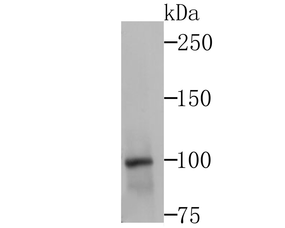
Western blot analysis of LAMP2a on different lysates with Rabbit anti-LAMP2a antibody (ET1601-24) at 1/5,000 dilution.
Lane 1: SK-MEL-28 cell lysate (20 µg/Lane)
Lane 2: HeLa cell lysate (20 µg/Lane)
Lane 3: JAR cell lysate (20 µg/Lane)
Lane 4: U-937 cell lysate (20 µg/Lane)
Lane 5: RAW264.7 cell lysate (15 µg/Lane)
Lane 6: NIH/3T3 cell lysate (15 µg/Lane)
Lane 7: PC-12 cell lysate (15 µg/Lane)
Lane 8: Mouse liver tissue lysate (30 µg/Lane)
Lane 9: Rat liver tissue lysate (30 µg/Lane)
Lane 10: Rat lung tissue lysate (30 µg/Lane)
Predicted band size: 45 kDa
Observed band size: 70-140 kDa
Exposure time: Lane 1-4: 2 minutes 37 seconds; Lane 5-10: 5 minutes;
4-20% SDS-PAGE gel.
Proteins were transferred to a PVDF membrane and blocked with 5% NFDM/TBST for 1 hour at room temperature. The primary antibody (ET1601-24) at 1/5,000 dilution was used in 5% NFDM/TBST at 4℃ overnight. Goat Anti-Rabbit IgG - HRP Secondary Antibody (HA1001) at 1:50,000 dilution was used for 1 hour at room temperature. -
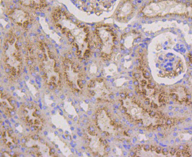
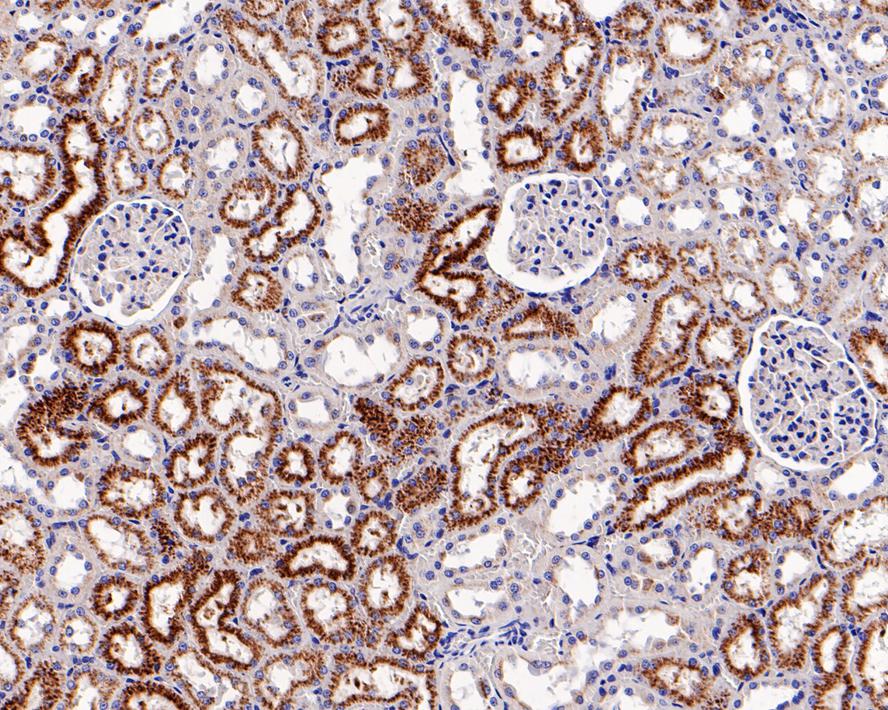
Immunohistochemical analysis of paraffin-embedded human kidney tissue with Rabbit anti-LAMP2a antibody (ET1601-24) at 1/500 dilution.
The section was pre-treated using heat mediated antigen retrieval with Tris-EDTA buffer (pH 9.0) for 20 minutes. The tissues were blocked in 1% BSA for 20 minutes at room temperature, washed with ddH2O and PBS, and then probed with the primary antibody (ET1601-24) at 1/500 dilution for 1 hour at room temperature. The detection was performed using an HRP conjugated compact polymer system. DAB was used as the chromogen. Tissues were counterstained with hematoxylin and mounted with DPX. -
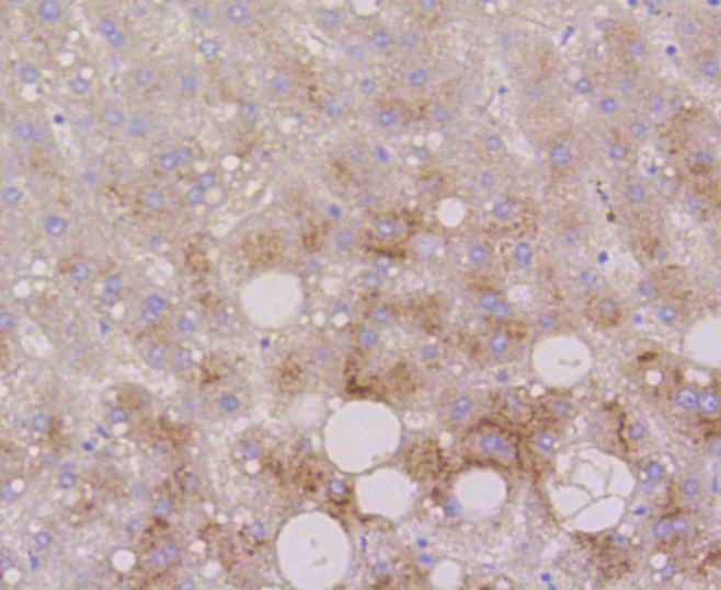
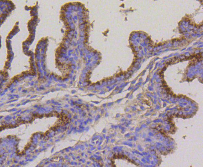
Immunohistochemical analysis of paraffin-embedded human liver tissue using anti-LAMP2a antibody. The section was pre-treated using heat mediated antigen retrieval with Tris-EDTA buffer (pH 8.0-8.4) for 20 minutes.The tissues were blocked in 5% BSA for 30 minutes at room temperature, washed with ddH2O and PBS, and then probed with the primary antibody (ET1601-24, 1/50) for 30 minutes at room temperature. The detection was performed using an HRP conjugated compact polymer system. DAB was used as the chromogen. Tissues were counterstained with hematoxylin and mounted with DPX.
-
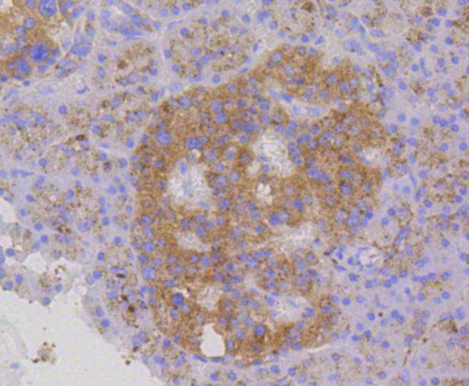
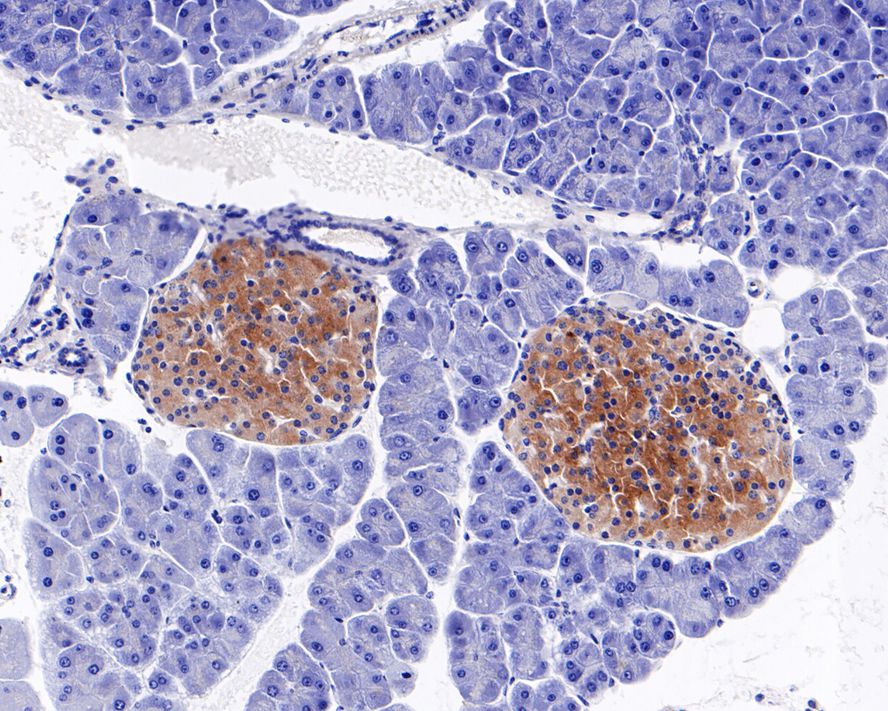
Immunohistochemical analysis of paraffin-embedded human pancreas tissue using anti-LAMP2a antibody. The section was pre-treated using heat mediated antigen retrieval with Tris-EDTA buffer (pH 8.0-8.4) for 20 minutes.The tissues were blocked in 5% BSA for 30 minutes at room temperature, washed with ddH2O and PBS, and then probed with the primary antibody (ET1601-24, 1/200) for 30 minutes at room temperature. The detection was performed using an HRP conjugated compact polymer system. DAB was used as the chromogen. Tissues were counterstained with hematoxylin and mounted with DPX.
-
Immunohistochemical analysis of paraffin-embedded mouse kidney tissue using anti-LAMP2a antibody. The section was pre-treated using heat mediated antigen retrieval with Tris-EDTA buffer (pH 8.0-8.4) for 20 minutes.The tissues were blocked in 5% BSA for 30 minutes at room temperature, washed with ddH2O and PBS, and then probed with the primary antibody (ET1601-24, 1/50) for 30 minutes at room temperature. The detection was performed using an HRP conjugated compact polymer system. DAB was used as the chromogen. Tissues were counterstained with hematoxylin and mounted with DPX.
-
Immunohistochemical analysis of paraffin-embedded mouse placenta tissue using anti-LAMP2a antibody. The section was pre-treated using heat mediated antigen retrieval with Tris-EDTA buffer (pH 8.0-8.4) for 20 minutes.The tissues were blocked in 5% BSA for 30 minutes at room temperature, washed with ddH2O and PBS, and then probed with the primary antibody (ET1601-24, 1/50) for 30 minutes at room temperature. The detection was performed using an HRP conjugated compact polymer system. DAB was used as the chromogen. Tissues were counterstained with hematoxylin and mounted with DPX.
-
Immunohistochemical analysis of paraffin-embedded mouse pancreas tissue with Rabbit anti-LAMP2a antibody (ET1601-24) at 1/500 dilution.
The section was pre-treated using heat mediated antigen retrieval with Tris-EDTA buffer (pH 9.0) for 20 minutes. The tissues were blocked in 1% BSA for 20 minutes at room temperature, washed with ddH2O and PBS, and then probed with the primary antibody (ET1601-24) at 1/500 dilution for 1 hour at room temperature. The detection was performed using an HRP conjugated compact polymer system. DAB was used as the chromogen. Tissues were counterstained with hematoxylin and mounted with DPX. -
Immunohistochemical analysis of paraffin-embedded rat kidney tissue with Rabbit anti-LAMP2a antibody (ET1601-24) at 1/500 dilution.
The section was pre-treated using heat mediated antigen retrieval with Tris-EDTA buffer (pH 9.0) for 20 minutes. The tissues were blocked in 1% BSA for 20 minutes at room temperature, washed with ddH2O and PBS, and then probed with the primary antibody (ET1601-24) at 1/500 dilution for 1 hour at room temperature. The detection was performed using an HRP conjugated compact polymer system. DAB was used as the chromogen. Tissues were counterstained with hematoxylin and mounted with DPX.
Please note: All products are "FOR RESEARCH USE ONLY AND ARE NOT INTENDED FOR DIAGNOSTIC OR THERAPEUTIC USE"
引文
-
Adenosine A2A receptor antagonist KW6002 protects against A53T mutant alpha-synuclein-induced brain damage and neuronal apoptosis in Parkinson's disease mice by restoring autophagic flux
Author: Hu Qidi,et al
PMID: 38157926
应用: IF-Tissue
反应种属: Mouse
发表时间: 2024 Jan
-
 Citation
-
Wei-Tong-Xin ameliorated cisplatin-induced mitophagy and apoptosis in gastric antral mucosa by activating the Nrf2/HO-1 pathway
Author:
PMID: 36806345

应用: WB
反应种属: Mouse
发表时间: 2023 May
-
Citation
-
Tumor cells induce LAMP2a expression in tumor-associated macrophage for cancer progression
Author: Yuquan Wei,Xiawei Wei
PMID: 30711520
应用: IP
反应种属: Mouse
发表时间: 2019 Feb
-
Citation
同靶点&同通路的产品