Collagen VI Recombinant Rabbit Monoclonal Antibody [SD83-03]


Catalog# ET1612-91
Collagen VI Recombinant Rabbit Monoclonal Antibody [SD83-03]
-
WB
-
IF-Cell
-
IF-Tissue
-
IHC-P
-
Human
-
Mouse
-
Rat
概述
产品名称
Collagen VI Recombinant Rabbit Monoclonal Antibody [SD83-03]
抗体类型
Recombinant Rabbit monoclonal Antibody
免疫原
Recombinant protein within Human Collagen VI alpha1 aa 17-255 / 1,028.
种属反应性
Human, Mouse, Rat
验证应用
WB, IF-Cell, IF-Tissue, IHC-P
分子量
Predicted band size: 109 kDa
阳性对照
NIH/3T3 cell lysate, human kidney tissue lysate, rat heart tissue lysate, rat kidney tissue lysate, Hela, A549, HepG2, RH-35, human lung tissue, human liver tissue, mouse colon tissue, human colon tissue, human skin tissue.
偶联
unconjugated
克隆号
SD83-03
RRID
产品特性
形态
Liquid
浓度
1ug/ul
存放说明
Store at +4℃ after thawing. Aliquot store at -20℃ or -80℃. Avoid repeated freeze / thaw cycles.
存储缓冲液
1*TBS (pH7.4), 0.05% BSA, 40% Glycerol. Preservative: 0.05% Sodium Azide.
亚型
IgG
纯化方式
Protein A affinity purified.
应用稀释度
-
WB
-
1:2,000
-
IF-Cell
-
1:100-1:500
-
IF-Tissue
-
1:100-1:500
-
IHC-P
-
1:50-1:200
发表文章中的应用
靶点
功能
Collagen VI (ColVI) is a type of collagen primarily associated with the extracellular matrix of skeletal muscle. ColVI maintains regularity in muscle function and stabilizes the cell membrane. It is synthesized by a complex, multistep pathway that leads to the formation of a unique network of linked microfilaments located in the extracellular matrix (ECM). ColVI plays a vital role in numerous cell types, including chondrocytes, neurons, myocytes, fibroblasts, and cardiomyocytes. ColVI molecules are made up of three alpha chains: α1(VI), α2(VI), and α3(VI). It is encoded by 6 genes: COL6A1, COL6A2, COL6A3, COL6A4, COL6A5, and COL6A6. The chain lengths of α1(VI) and α2(VI) are about 1,000 amino acids. The chain length of α3(VI) is roughly a third larger than those of α1(VI) and α2(VI), and it consists of several spliced variants within the range of 2,500 to 3,100 amino acids. The first two alpha chains subunits of ColVI have a molecular weight of 140-150 KDa and the third polypeptide chain is larger with a molecular weight of 250-300kDa. ColVI is also found in the skin, lungs, blood vessels, cornea and intervertebral disc. It also forms part of the peripheral nerves, brain, myocardium and adipose tissue.
背景文献
1. Kristofik N et al. Impaired von Willebrand factor adhesion and platelet response in thrombospondin-2 knockout mice. Blood 128:1642-50 (2016).
2. Okawa S et al. Lipopolysaccharide induces expression of collagen VI in the rat lung. J Toxicol Pathol 28:37-41 (2015).
序列相似性
Belongs to the type VI collagen family.
翻译后修饰
Prolines at the third position of the tripeptide repeating unit (G-X-Y) are hydroxylated in some or all of the chains.
亚细胞定位
Extracellular matrix.
UNIPROT #
别名
Alpha 1 (VI) chain (61 AA) antibody
CO6A1_HUMAN antibody
COL6A1 antibody
Collagen alpha-1(VI) chain antibody
Collagen type VI alpha 1 antibody
Collagen VI alpha 1 polypeptide antibody
CollagenVI antibody
Human mRNA for collagen VI alpha 2 C terminal globular domain antibody
OPLL antibody
PP3610 antibody
图片
-
Western blot analysis of Collagen VI on different lysates with Rabbit anti-Collagen VI antibody (ET1612-91) at 1/2,000 dilution.
Lane 1: NIH/3T3 cell lysate (15 µg/Lane)
Lane 2: Human kidney tissue lysate (20 µg/Lane)
Lane 3: Rat heart tissue lysate (20 µg/Lane)
Lane 4: Rat kidney tissue lysate (20 µg/Lane)
Predicted band size: 109 kDa
Observed band size: 145 kDa
Exposure time: 1 minute 30 seconds;
4-20% SDS-PAGE gel.
Proteins were transferred to a PVDF membrane and blocked with 5% NFDM/TBST for 1 hour at room temperature. The primary antibody (ET1612-91) at 1/2,000 dilution was used in 5% NFDM/TBST at 4℃ overnight. Goat Anti-Rabbit IgG - HRP Secondary Antibody (HA1001) at 1/50,000 dilution was used for 1 hour at room temperature. -
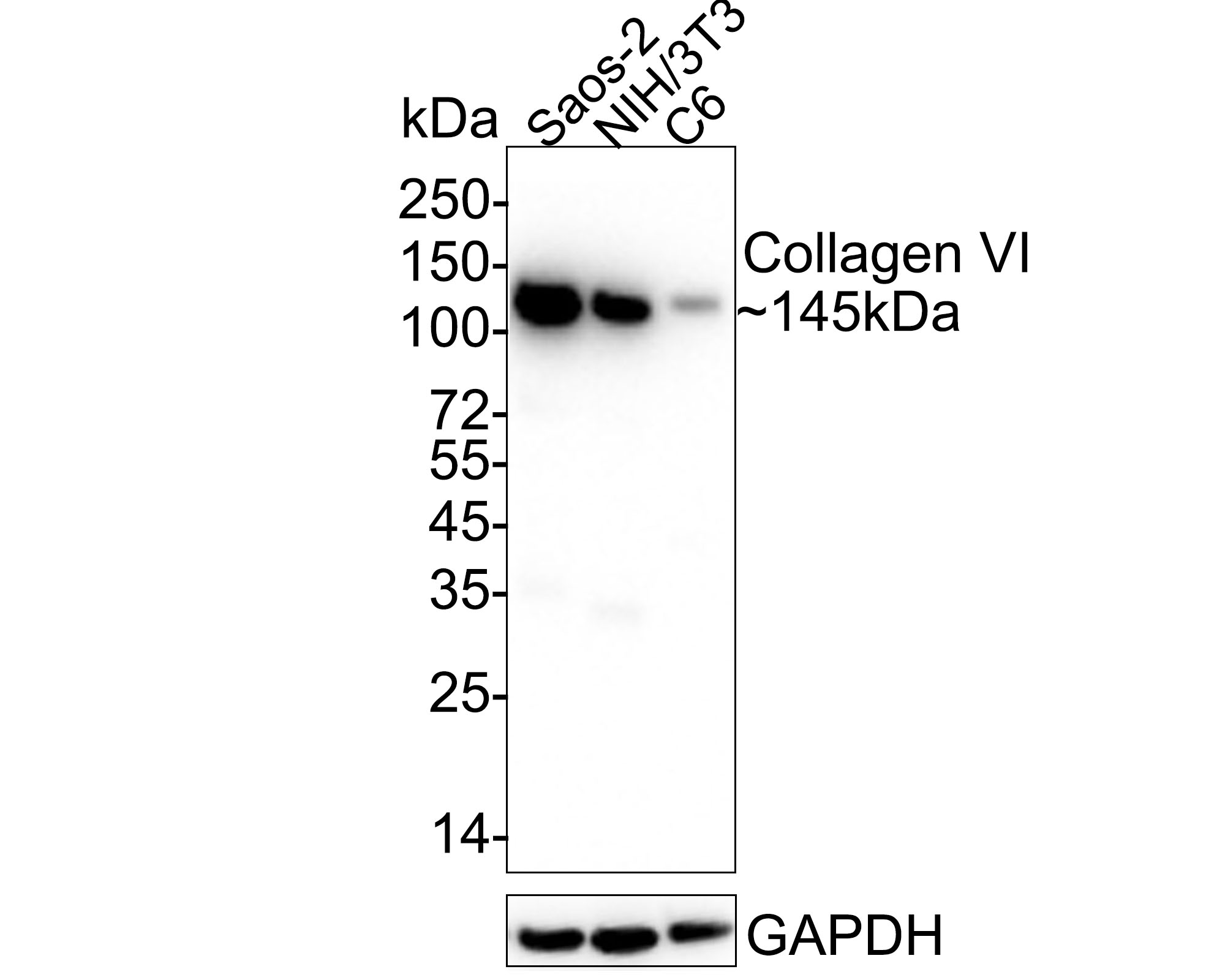
Western blot analysis of Collagen VI on different lysates with Rabbit anti-Collagen VI antibody (ET1612-91) at 1/1,000 dilution.
Lane 1: Saos-2 cell lysate
Lane 2: NIH/3T3 cell lysate
Lane 3: C6 cell lysate
Lysates/proteins at 20 µg/Lane.
Predicted band size: 145 kDa
Observed band size: 145 kDa
Exposure time: 6 seconds;
4-20% SDS-PAGE gel.
Proteins were transferred to a PVDF membrane and blocked with 5% NFDM/TBST for 1 hour at room temperature. The primary antibody (ET1612-91) at 1/1,000 dilution was used in 5% NFDM/TBST at 4℃ overnight. Goat Anti-Rabbit IgG - HRP Secondary Antibody (HA1001) at 1/50,000 dilution was used for 1 hour at room temperature. -
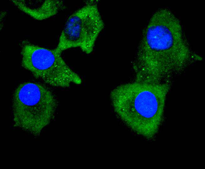
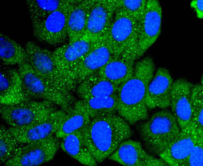
ICC staining of Collagen VI in Hela cells (green). Formalin fixed cells were permeabilized with 0.1% Triton X-100 in TBS for 10 minutes at room temperature and blocked with 10% negative goat serum for 15 minutes at room temperature. Cells were probed with the primary antibody (ET1612-91, 1/50) for 1 hour at room temperature, washed with PBS. Alexa Fluor®488 conjugate-Goat anti-Rabbit IgG was used as the secondary antibody at 1/1,000 dilution. The nuclear counter stain is DAPI (blue).
-
ICC staining of Collagen VI in A549 cells (green). Formalin fixed cells were permeabilized with 0.1% Triton X-100 in TBS for 10 minutes at room temperature and blocked with 10% negative goat serum for 15 minutes at room temperature. Cells were probed with the primary antibody (ET1612-91, 1/50) for 1 hour at room temperature, washed with PBS. Alexa Fluor®488 conjugate-Goat anti-Rabbit IgG was used as the secondary antibody at 1/1,000 dilution. The nuclear counter stain is DAPI (blue).
-
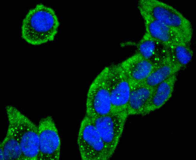
ICC staining of Collagen VI in HepG2 cells (green). Formalin fixed cells were permeabilized with 0.1% Triton X-100 in TBS for 10 minutes at room temperature and blocked with 10% negative goat serum for 15 minutes at room temperature. Cells were probed with the primary antibody (ET1612-91, 1/50) for 1 hour at room temperature, washed with PBS. Alexa Fluor®488 conjugate-Goat anti-Rabbit IgG was used as the secondary antibody at 1/1,000 dilution. The nuclear counter stain is DAPI (blue).
-
ICC staining of Collagen VI in RH-35 cells (green). Formalin fixed cells were permeabilized with 0.1% Triton X-100 in TBS for 10 minutes at room temperature and blocked with 10% negative goat serum for 15 minutes at room temperature. Cells were probed with the primary antibody (ET1612-91, 1/50) for 1 hour at room temperature, washed with PBS. Alexa Fluor®488 conjugate-Goat anti-Rabbit IgG was used as the secondary antibody at 1/1,000 dilution. The nuclear counter stain is DAPI (blue).
-
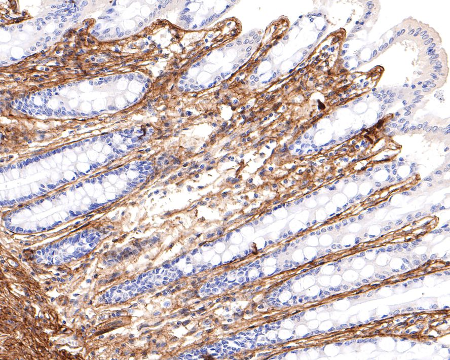
Immunohistochemical analysis of paraffin-embedded human lung tissue using anti-Collagen VI antibody. The section was pre-treated using heat mediated antigen retrieval with Tris-EDTA buffer (pH 9.0) for 20 minutes.The tissues were blocked in 1% BSA for 30 minutes at room temperature, washed with ddH2O and PBS, and then probed with the primary antibody (ET1612-91, 1/50) for 30 minutes at room temperature. The detection was performed using an HRP conjugated compact polymer system. DAB was used as the chromogen. Tissues were counterstained with hematoxylin and mounted with DPX.
-
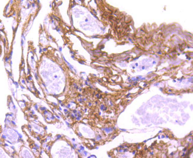
Immunohistochemical analysis of paraffin-embedded human liver tissue with Rabbit anti-Collagen VI antibody (ET1612-91) at 1/400 dilution.
The section was pre-treated using heat mediated antigen retrieval with Tris-EDTA buffer (pH 9.0) for 20 minutes. The tissues were blocked in 1% BSA for 20 minutes at room temperature, washed with ddH2O and PBS, and then probed with the primary antibody (ET1612-91) at 1/400 dilution for 1 hour at room temperature. The detection was performed using an HRP conjugated compact polymer system. DAB was used as the chromogen. Tissues were counterstained with hematoxylin and mounted with DPX. -
Immunohistochemical analysis of paraffin-embedded mouse colon tissue using anti-Collagen VI antibody. The section was pre-treated using heat mediated antigen retrieval with Tris-EDTA buffer (pH 9.0) for 20 minutes.The tissues were blocked in 1% BSA for 30 minutes at room temperature, washed with ddH2O and PBS, and then probed with the primary antibody (ET1612-91, 1/50) for 30 minutes at room temperature. The detection was performed using an HRP conjugated compact polymer system. DAB was used as the chromogen. Tissues were counterstained with hematoxylin and mounted with DPX.
-
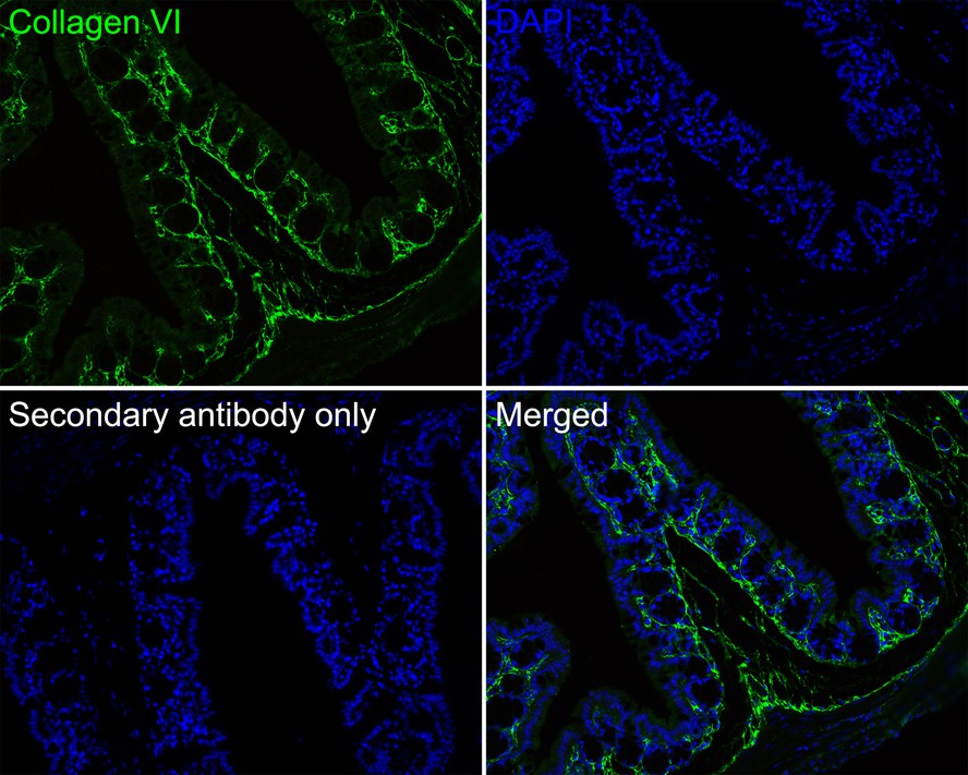
Immunohistochemical analysis of paraffin-embedded human colon tissue with Rabbit anti-Collagen VI antibody (ET1612-91) at 1/400 dilution.
The section was pre-treated using heat mediated antigen retrieval with Tris-EDTA buffer (pH 9.0) for 20 minutes. The tissues were blocked in 1% BSA for 20 minutes at room temperature, washed with ddH2O and PBS, and then probed with the primary antibody (ET1612-91) at 1/400 dilution for 1 hour at room temperature. The detection was performed using an HRP conjugated compact polymer system. DAB was used as the chromogen. Tissues were counterstained with hematoxylin and mounted with DPX. -
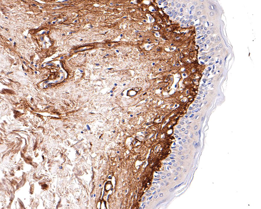
Immunohistochemical analysis of paraffin-embedded human skin tissue with Rabbit anti-Collagen VI antibody (ET1612-91) at 1/400 dilution.
The section was pre-treated using heat mediated antigen retrieval with Tris-EDTA buffer (pH 9.0) for 20 minutes. The tissues were blocked in 1% BSA for 20 minutes at room temperature, washed with ddH2O and PBS, and then probed with the primary antibody (ET1612-91) at 1/400 dilution for 1 hour at room temperature. The detection was performed using an HRP conjugated compact polymer system. DAB was used as the chromogen. Tissues were counterstained with hematoxylin and mounted with DPX. -
Immunofluorescence analysis of paraffin-embedded human liver tissue labeling Collagen VI with Rabbit anti-Collagen VI antibody (ET1612-91) at 1/100 dilution.
The section was pre-treated using heat mediated antigen retrieval with Tris-EDTA buffer (pH 9.0) for 20 minutes. The tissues were blocked in 10% negative goat serum for 1 hour at room temperature, washed with PBS, and then probed with the primary antibody (ET1612-91, green) at 1/100 dilution overnight at 4 ℃, washed with PBS.
Goat Anti-Rabbit IgG H&L (iFluor™ 488, HA1121) was used as the secondary antibody at 1/1,000 dilution. Nuclei were counterstained with DAPI (blue). -
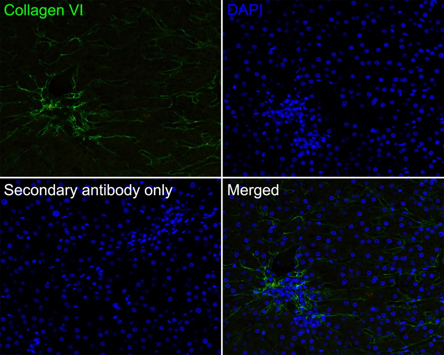
Immunofluorescence analysis of paraffin-embedded mouse colon tissue labeling Collagen VI with Rabbit anti-Collagen VI antibody (ET1612-91) at 1/100 dilution.
The section was pre-treated using heat mediated antigen retrieval with Tris-EDTA buffer (pH 9.0) for 20 minutes. The tissues were blocked in 10% negative goat serum for 1 hour at room temperature, washed with PBS, and then probed with the primary antibody (ET1612-91, green) at 1/100 dilution overnight at 4 ℃, washed with PBS.
Goat Anti-Rabbit IgG H&L (iFluor™ 488, HA1121) was used as the secondary antibody at 1/1,000 dilution. Nuclei were counterstained with DAPI (blue). -
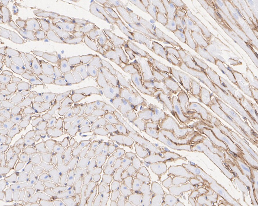
Immunohistochemical analysis of paraffin-embedded rat heart tissue with Rabbit anti-Collagen VI antibody (ET1612-91) at 1/1,000 dilution.
The section was pre-treated using heat mediated antigen retrieval with Tris-EDTA buffer (pH 9.0) for 20 minutes. The tissues were blocked in 1% BSA for 20 minutes at room temperature, washed with ddH2O and PBS, and then probed with the primary antibody (ET1612-91) at 1/1,000 dilution for 1 hour at room temperature. The detection was performed using an HRP conjugated compact polymer system. DAB was used as the chromogen. Tissues were counterstained with hematoxylin and mounted with DPX. -
Immunohistochemical analysis of paraffin-embedded rat kidney tissue with Rabbit anti-Collagen VI antibody (ET1612-91) at 1/1,000 dilution.
The section was pre-treated using heat mediated antigen retrieval with Tris-EDTA buffer (pH 9.0) for 20 minutes. The tissues were blocked in 1% BSA for 20 minutes at room temperature, washed with ddH2O and PBS, and then probed with the primary antibody (ET1612-91) at 1/1,000 dilution for 1 hour at room temperature. The detection was performed using an HRP conjugated compact polymer system. DAB was used as the chromogen. Tissues were counterstained with hematoxylin and mounted with DPX. -
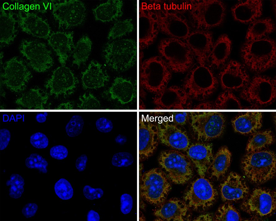
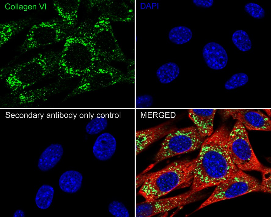
Immunocytochemistry analysis of NIH/3T3 cells labeling Collagen VI with Rabbit anti-Collagen VI antibody (ET1612-91) at 1/100 dilution.
Cells were fixed in 4% paraformaldehyde for 20 minutes at room temperature, permeabilized with 0.1% Triton X-100 in PBS for 5 minutes at room temperature, then blocked with 1% BSA in 10% negative goat serum for 1 hour at room temperature. Cells were then incubated with Rabbit anti-Collagen VI antibody (ET1612-91) at 1/100 dilution in 1% BSA in PBST overnight at 4 ℃. Goat Anti-Rabbit IgG H&L (iFluor™ 488, HA1121) was used as the secondary antibody at 1/1,000 dilution. PBS instead of the primary antibody was used as the secondary antibody only control. Nuclear DNA was labelled in blue with DAPI.
Beta tubulin (M1305-2, red) was stained at 1/100 dilution overnight at +4℃. Goat Anti-Mouse IgG H&L (iFluor™ 594, HA1126) was used as the secondary antibody at 1/1,000 dilution.
Please note: All products are "FOR RESEARCH USE ONLY AND ARE NOT INTENDED FOR DIAGNOSTIC OR THERAPEUTIC USE"
引文
-
TGF-β1-induced collagen promotes chicken ovarian follicle development via an intercellular cooperative pattern
Author: Zhou, S., Ma, Y., Yao, J., Zhao, A., Xie, C., Mi, Y., & Zhang, C.
PMID: 33675281

应用: WB
反应种属: Hen
发表时间: 2021 Jun
-
 Citation
-
Transcriptome profiling analysis of underlying regulation of growing follicle development in the chicken
Author: Caiqiao Zhang
PMID: 32475419
应用: IHC,WB
反应种属: chicken
发表时间: 2020 Jun
-
Citation
同靶点&同通路的产品