CD133 Mouse Monoclonal Antibody [A8C3]


Catalog# HA601024
CD133 Mouse Monoclonal Antibody [A8C3]
-
WB
-
IHC-P
-
FC
-
Human
概述
产品名称
CD133 Mouse Monoclonal Antibody [A8C3]
抗体类型
Mouse Monoclonal Antibody
免疫原
Recombinant protein within human CD133 aa 151-400/865.
种属反应性
Human
验证应用
WB, IHC-P, FC
分子量
Predicted band size: 97 kDa
阳性对照
NCCIT cell lysates, human kidney tissue lysates, HT-29 cell lysates, human colon carcinoma tissue, human breast tissue, human kidney tissue, NCCIT.
偶联
unconjugated
克隆号
A8C3
RRID
产品特性
形态
Liquid
浓度
2ug/ul
存放说明
Store at +4℃ after thawing. Aliquot store at -20℃. Avoid repeated freeze / thaw cycles.
存储缓冲液
PBS (pH7.4), 0.1% BSA, 40% Glycerol. Preservative: 0.05% Sodium Azide.
亚型
IgG1
纯化方式
Protein G affinity purified.
应用稀释度
-
WB
-
1:500
-
IHC-P
-
1:600
-
FC
-
1:500-1:1,000
靶点
功能
CD133 antigen, also known as prominin-1, is a glycoprotein that in humans is encoded by the PROM1 gene. It is a member of pentaspan transmembrane glycoproteins, which specifically localize to cellular protrusions. When embedded in the cell membrane, the membrane topology of prominin-1 is such that the N-terminus extends into the extracellular space and the C-terminus resides in the intracellular compartment. The protein consists of five transmembrane segments, with the first and second segments and the third and fourth segments connected by intracellular loops while the second and third as well as fourth and fifth transmembrane segments are connected by extracellular loops. CD133 is the most commonly used marker for isolation of cancer stem cell (CSC) population from different tumors, mainly from various gliomas and carcinomas. CD133+ melanoma cells are considered a subpopulation of CSC and play a critical role in recurrence. Moreover, CD133+ melanoma cells are immunogenic and can be used as an antimelanoma vaccination. In mice the vaccination with CD133+ melanoma cells mediated strong anti-tumor activity that resulted in the eradication of parental melanoma cells. In addition, it has also been shown that CD133+ melanoma cells preferentially express the RNA helicase DDX3X . As DDX3X also is an immunogenic protein, the same anti-melanoma vaccination strategy can be employed to give therapeutic antitumor immunity in mice.
背景文献
1. Kim MY et al. Accumulation of low-dose BIX01294 promotes metastatic potential of U251 glioblastoma cells. Oncol Lett 13:1767-1774 (2017).
2. Xi G et al. Targeting CD133 improves chemotherapeutic efficacy of recurrent pediatric pilocytic astrocytoma following prolonged chemotherapy. Mol Cancer 16:21 (2017).
亚细胞定位
Endoplasmic reticulum. Plasma membrane. Cell projection.
UNIPROT #
别名
AC133 antibody
Antigen AC133 antibody
CD133 antibody
CORD12 antibody
Hematopoietic stem cell antigen antibody
hProminin antibody
MCDR2 antibody
MSTP061 antibody
OTTHUMP00000217744 antibody
OTTHUMP00000217745 antibody
展开图片
-
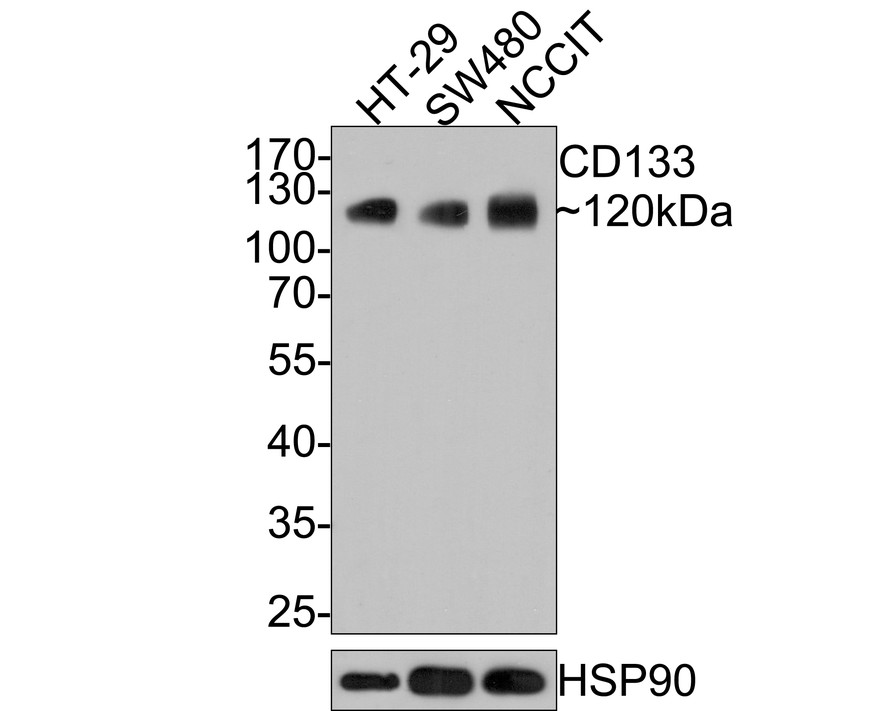
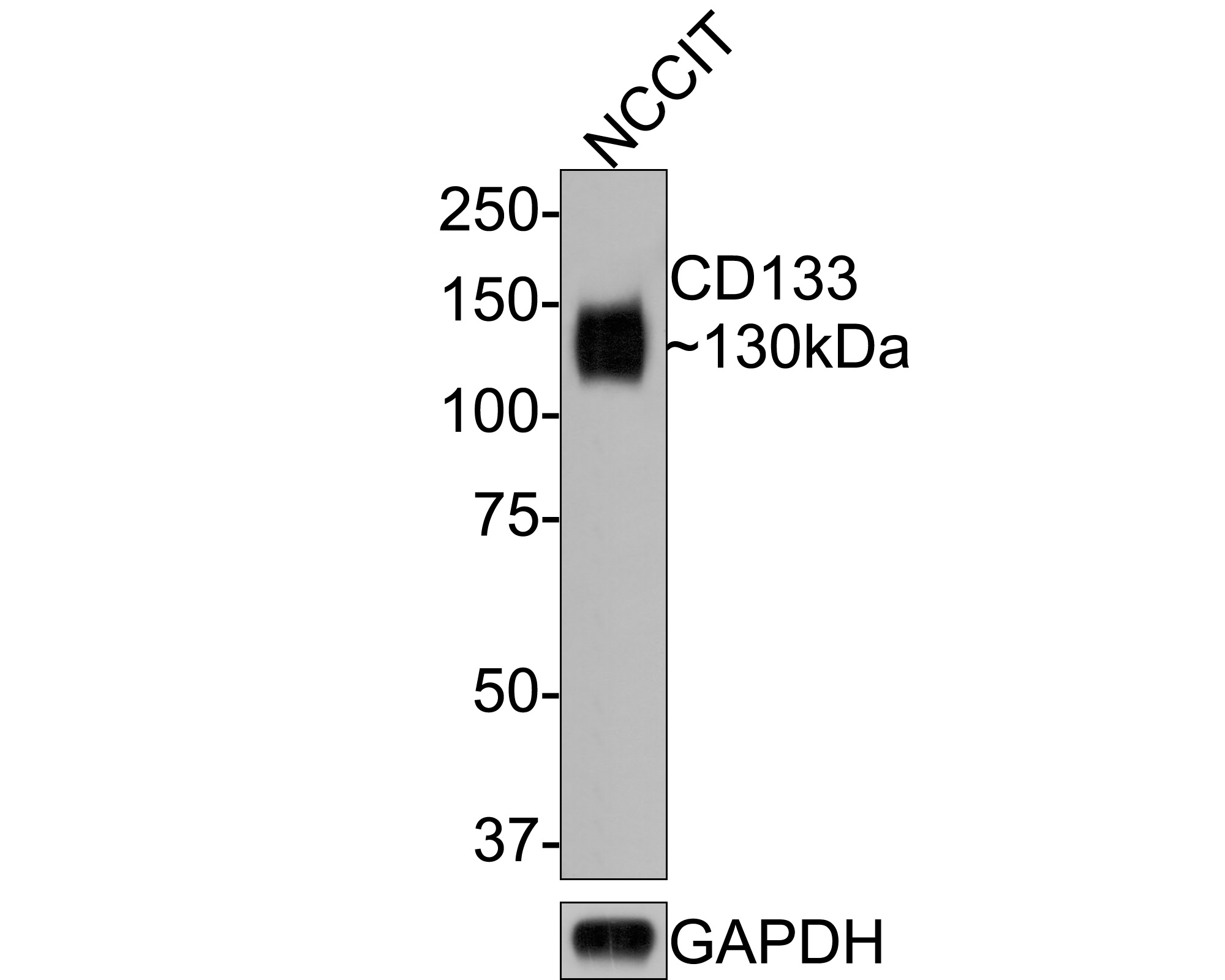
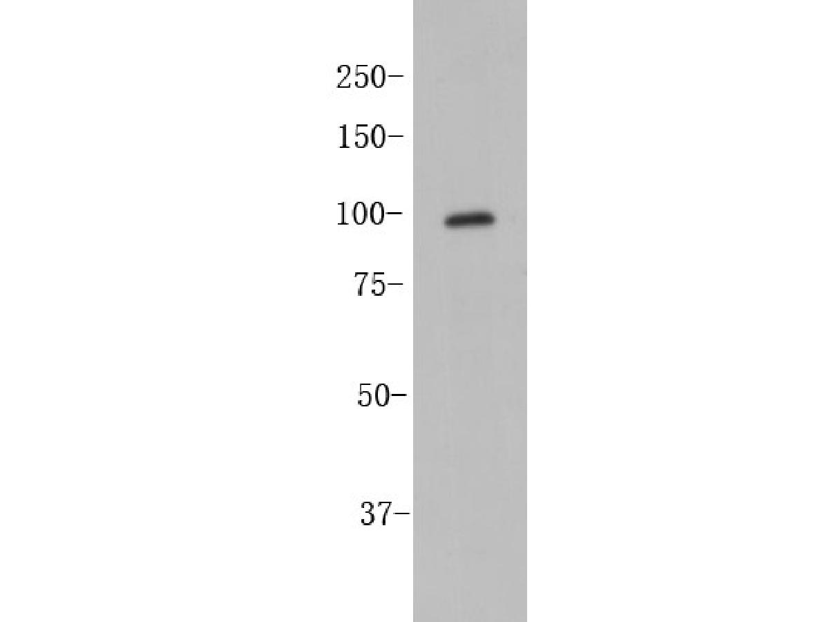
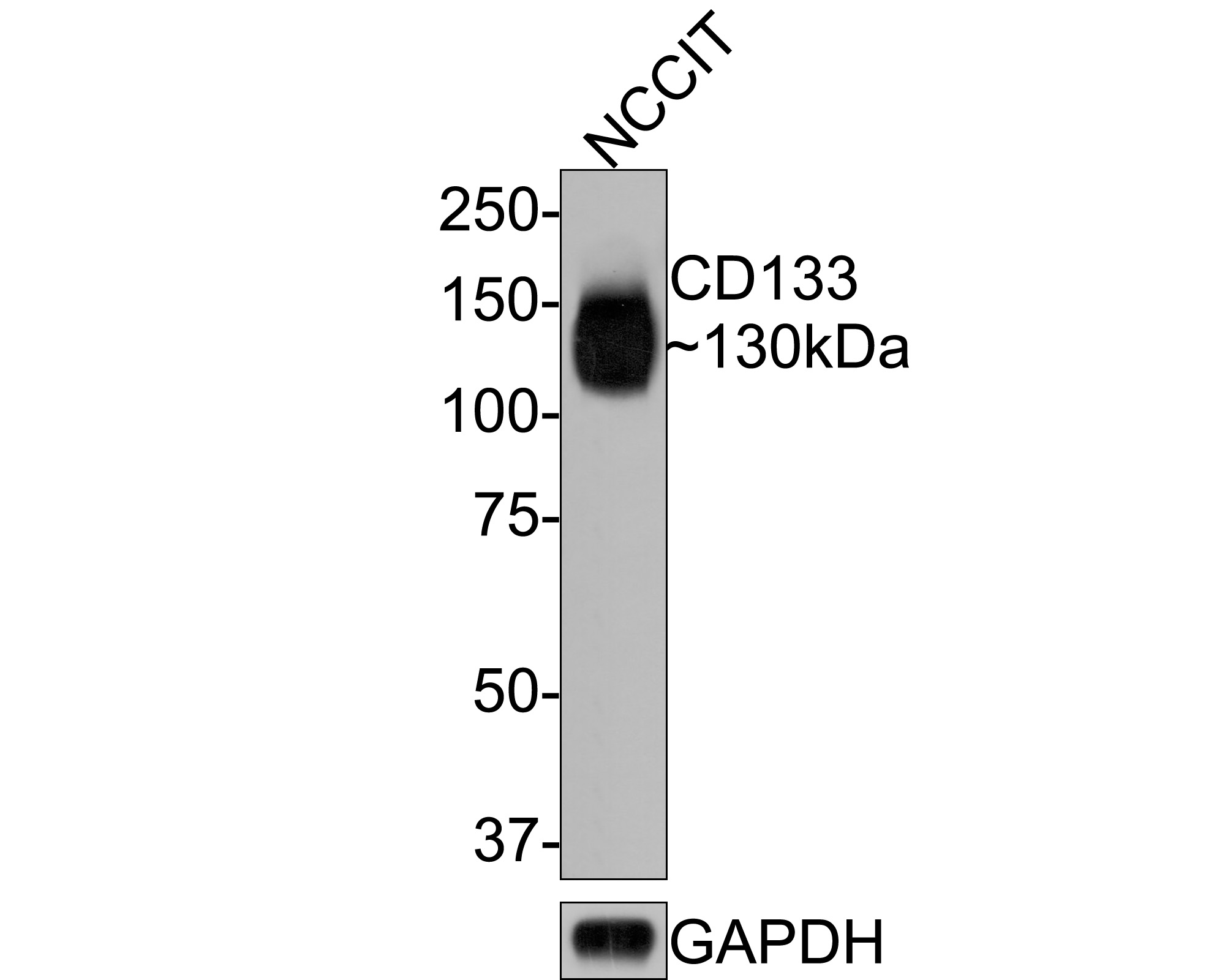
Western blot analysis of CD133 on NCCIT cell lysates with Mouse anti-CD133 antibody (HA601024) at 1/500 dilution.
Lysates/proteins at 10 µg/Lane.
Predicted band size: 97 kDa
Observed band size: 130 kDa
Exposure time: 2 minutes;
8% SDS-PAGE gel.
Proteins were transferred to a PVDF membrane and blocked with 5% NFDM/TBST for 1 hour at room temperature. The primary antibody (HA601024) at 1/500 dilution was used in 5% NFDM/TBST at room temperature for 2 hours. Goat Anti-Mouse IgG - HRP Secondary Antibody (HA1006) at 1:100,000 dilution was used for 1 hour at room temperature. -
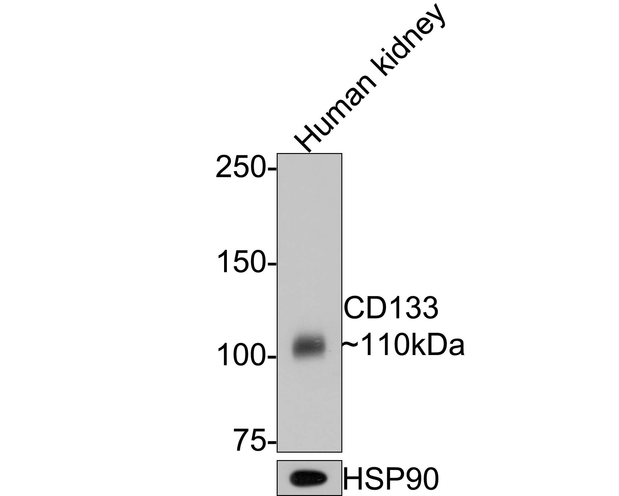
Western blot analysis of CD133 on human kidney tissue lysates with Mouse anti-CD133 antibody (HA601024) at 1/500 dilution.
Lysates/proteins at 20 µg/Lane.
Predicted band size: 97 kDa
Observed band size: 110 kDa
Exposure time: 30 seconds;
6% SDS-PAGE gel.
Proteins were transferred to a PVDF membrane and blocked with 5% NFDM/TBST for 1 hour at room temperature. The primary antibody (HA601024) at 1/500 dilution was used in 5% NFDM/TBST at room temperature for 2 hours. Goat Anti-Mouse IgG - HRP Secondary Antibody (HA1006) at 1:100,000 dilution was used for 1 hour at room temperature. -
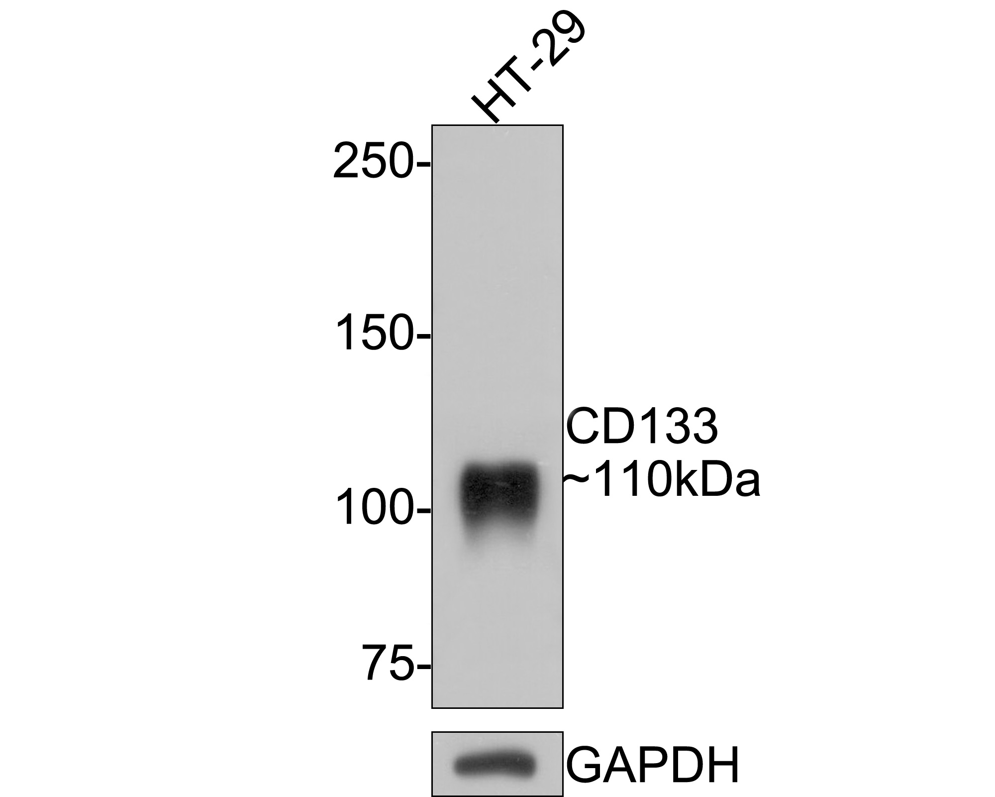
Western blot analysis of CD133 on HT-29 cell lysates with Mouse anti-CD133 antibody (HA601024) at 1/500 dilution.
Lysates/proteins at 10 µg/Lane.
Predicted band size: 97 kDa
Observed band size: 110 kDa
Exposure time: 1 minute;
6% SDS-PAGE gel.
Proteins were transferred to a PVDF membrane and blocked with 5% NFDM/TBST for 1 hour at room temperature. The primary antibody (HA601024) at 1/500 dilution was used in 5% NFDM/TBST at room temperature for 2 hours. Goat Anti-Mouse IgG - HRP Secondary Antibody (HA1006) at 1:100,000 dilution was used for 1 hour at room temperature. -
Immunohistochemical analysis of paraffin-embedded human colon carcinoma tissue with Mouse anti-CD133 antibody (HA601024) at 1/600 dilution.
The section was pre-treated using heat mediated antigen retrieval with Tris-EDTA buffer (pH 9.0) for 20 minutes. The tissues were blocked in 1% BSA for 20 minutes at room temperature, washed with ddH2O and PBS, and then probed with the primary antibody (HA601024) at 1/600 dilution for 1 hour at room temperature. The detection was performed using an HRP conjugated compact polymer system. DAB was used as the chromogen. Tissues were counterstained with hematoxylin and mounted with DPX. -
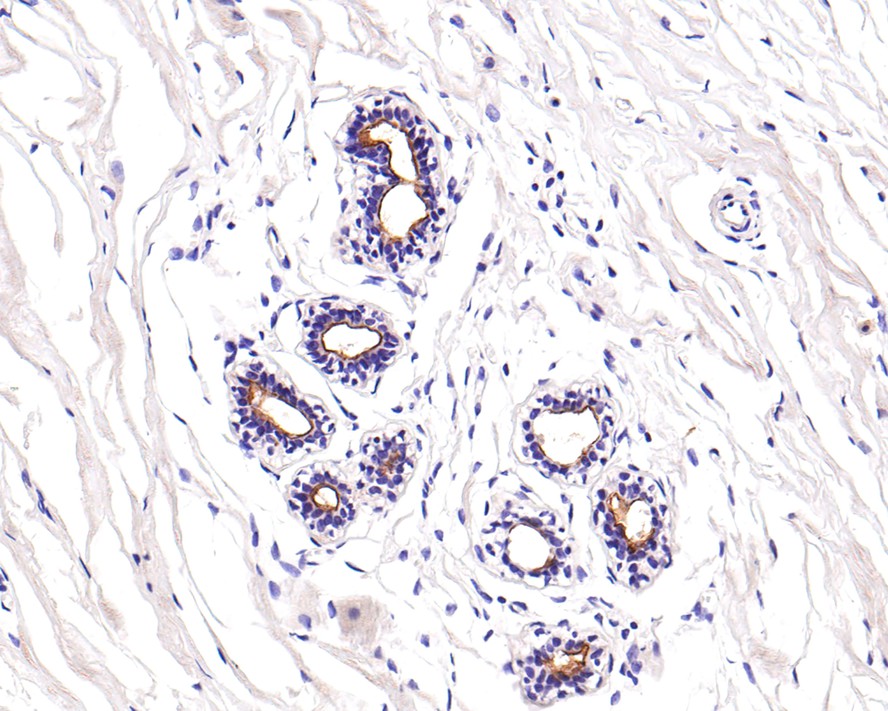
Immunohistochemical analysis of paraffin-embedded human breast tissue with Mouse anti-CD133 antibody (HA601024) at 1/600 dilution.
The section was pre-treated using heat mediated antigen retrieval with Tris-EDTA buffer (pH 9.0) for 20 minutes. The tissues were blocked in 1% BSA for 20 minutes at room temperature, washed with ddH2O and PBS, and then probed with the primary antibody (HA601024) at 1/600 dilution for 1 hour at room temperature. The detection was performed using an HRP conjugated compact polymer system. DAB was used as the chromogen. Tissues were counterstained with hematoxylin and mounted with DPX. -
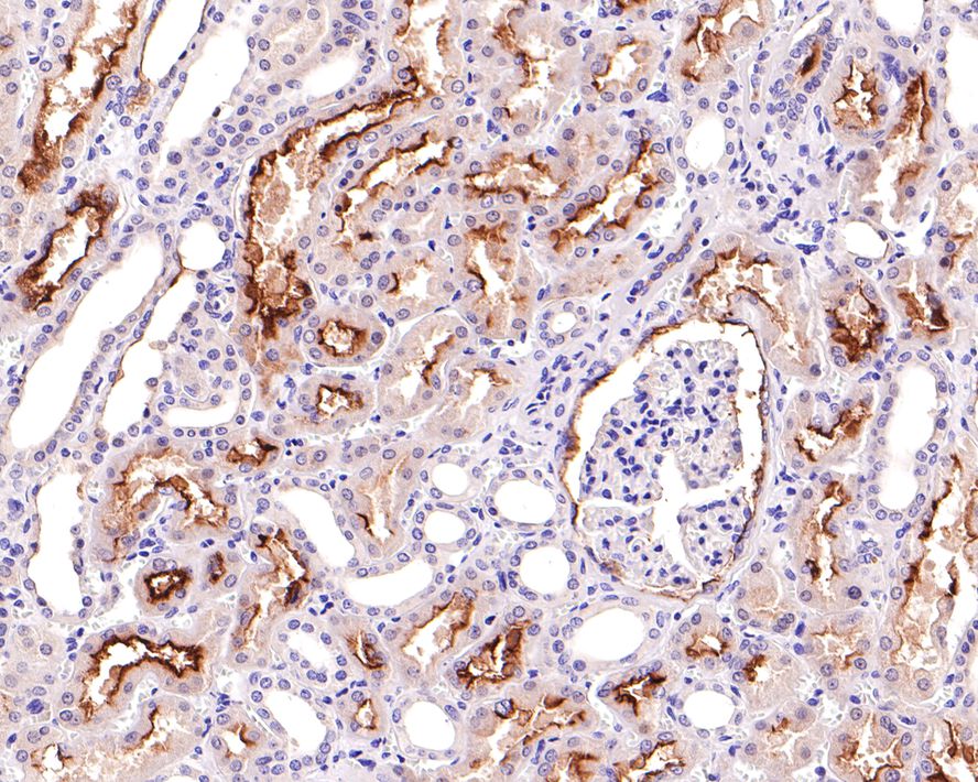
Immunohistochemical analysis of paraffin-embedded human kidney tissue with Mouse anti-CD133 antibody (HA601024) at 1/600 dilution.
The section was pre-treated using heat mediated antigen retrieval with Tris-EDTA buffer (pH 9.0) for 20 minutes. The tissues were blocked in 1% BSA for 20 minutes at room temperature, washed with ddH2O and PBS, and then probed with the primary antibody (HA601024) at 1/600 dilution for 1 hour at room temperature. The detection was performed using an HRP conjugated compact polymer system. DAB was used as the chromogen. Tissues were counterstained with hematoxylin and mounted with DPX. -
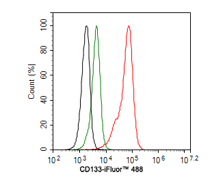
Flow cytometric analysis of NCCIT cells labeling CD133.
Cells were fixed and permeabilized. Then stained with the primary antibody (HA601024, 1ug/ml) (red) compared with Mouse IgG Isotype Control (green). After incubation of the primary antibody at +4℃ for an hour, the cells were stained with a iFluor™ 488 conjugate-Goat anti-Mouse IgG Secondary antibody at 1/1,000 dilution for 30 minutes at +4℃. Unlabelled sample was used as a control (cells without incubation with primary antibody; black).
Please note: All products are "FOR RESEARCH USE ONLY AND ARE NOT INTENDED FOR DIAGNOSTIC OR THERAPEUTIC USE"
同靶点&同通路的产品
CD133 Rabbit Polyclonal Antibody
Application: WB,FC
Reactivity: Human
Conjugate: unconjugated

CD133 Rabbit Polyclonal Antibody
Application: WB,FC
Reactivity: Human,Mouse,Rat
Conjugate: unconjugated
CD133 Rabbit Polyclonal Antibody
Application: WB,IHC-P,FC
Reactivity: Human
Conjugate: unconjugated
CD133 Mouse Monoclonal Antibody [A8C2]
Application: WB,IHC-P,FC
Reactivity: Human
Conjugate: unconjugated