iFluor™ 488 Conjugated E-Cadherin Recombinant Rabbit Monoclonal Antibody [SY0287]


Catalog# HA720159F
iFluor™ 488 Conjugated E-Cadherin Recombinant Rabbit Monoclonal Antibody [SY0287]
-
IF-Cell
-
FC
-
Human
概述
产品名称
iFluor™ 488 Conjugated E-Cadherin Recombinant Rabbit Monoclonal Antibody [SY0287]
抗体类型
Recombinant Rabbit monoclonal Antibody
免疫原
Synthetic peptide within Human E-Cadherin aa 591-640 / 882.
种属反应性
Human
验证应用
IF-Cell, FC
分子量
Predicted band size: 97 kDa
阳性对照
MCF-7, A431.
偶联
iFluor™ 488
克隆号
SY0287
RRID
产品特性
形态
Liquid
浓度
1ug/ul
存放说明
Store at +4℃ after thawing. Aliquot store at -20℃. Avoid repeated freeze / thaw cycles.
存储缓冲液
Preservative: 0.02% Sodium azide Constituents: 30% Glycerol, 1% BSA, 68.98% PBS
亚型
IgG
纯化方式
Protein A affinity purified.
应用稀释度
-
IF-Cell
-
1:100
-
FC
-
1:50-1:1,000
靶点
功能
Cadherins comprise a family of Ca2+-dependent adhesion molecules that function to mediate cell-cell binding critical to the maintenance of tissue structure and morphogenesis. Members of this family of adhesion proteins include rat cadherin K (and its human homolog, cadherin-6), R-cadherin, B-cadherin, E/P cadherin and cadherin-5. The classical cadherins, E-, N- and P-cadherin, consist of large extracellular domains characterized by a series of five homologous NH2 terminal repeats. The most distal of these cadherins is thought to be responsible for binding specificity, transmembrane domains and carboxy terminal intracellular domains. The relatively short intracellular domains interact with a variety of cytoplasmic proteins, such as β-catenin, to regulate cadherin function.
背景文献
1. Su B et al. Diallyl disulfide suppresses epithelial-mesenchymal transition, invasion and proliferation by downregulation of LIMK1 in gastric cancer. Oncotarget 7:10498-512 (2016).
2. Schmidt TP et al. Identification of E-cadherin signature motifs functioning as cleavage sites for Helicobacter pylori HtrA. Sci Rep 6:23264 (2016).
亚细胞定位
Endosome, Cell membrane, trans-Golgi network, adherens junction.
UNIPROT
别名
Arc 1 antibody
CADH1_HUMAN antibody
Cadherin 1 antibody
cadherin 1 type 1 E-cadherin antibody
Cadherin1 antibody
CAM 120/80 antibody
CD 324 antibody
CD324 antibody
CD324 antigen antibody
cdh1 antibody
展开图片
-
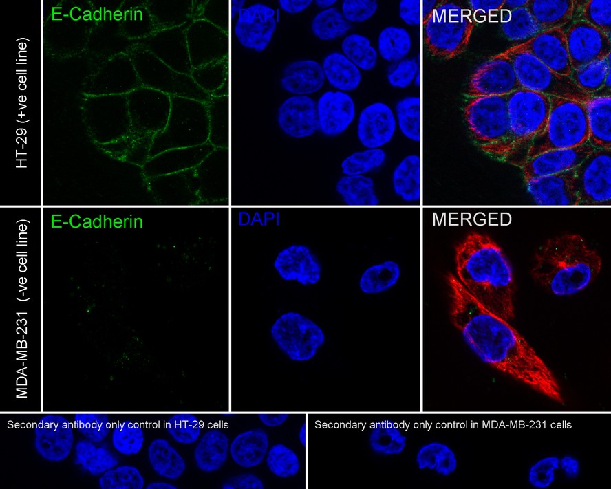
Immunocytochemistry analysis of MCF-7 cells labeling E-Cadherin with Rabbit anti-E-Cadherin antibody (HA720159F) at 1/100 dilution.
Cells were fixed in 4% paraformaldehyde for 10 minutes, permeabilized with 0.1% Triton X-100 in PBS for 15 minutes, and then blocked with 2% normal goat serum for 1 hour at 37 ℃. Cells were then incubated with Rabbit anti-E-Cadherin antibody (HA720159F) at 1/100 dilution in 2% normal goat serum overnight at 4 ℃. Nuclear DNA was labelled in blue with DAPI.
Beta tubulin (M1305-2, red) was stained at 1/200 dilution overnight at +4℃. Goat Anti-Mouse IgG H&L (iFluor™ 594, HA1126) were used as the secondary antibody at 1/800 dilution. -
Flow cytometric analysis of A431 cells labeling E-Cadherin.
Cells were washed twice with cold PBS and resuspend. Then incubated for 30 minutes at +4℃ with E-Cadherin (HA720159F, red, 1ug/ml) and Rabbit IgG Isotype Control (iFluor™ 488, green, 1ug/ml). Unlabelled sample was used as a control (cells without incubation with primary antibody; black). -
Flow cytometric analysis of MCF-7 cells labeling E-Cadherin.
Cells were washed twice with cold PBS and resuspend. Then incubated for 30 minutes at +4℃ with E-Cadherin (HA720159F, red, 10ug/ml) and Rabbit IgG Isotype Control (iFluor™ 488, green, 10ug/ml). Unlabelled sample was used as a control (cells without incubation with primary antibody; black).
请注意: All products are "FOR RESEARCH USE ONLY AND ARE NOT INTENDED FOR DIAGNOSTIC OR THERAPEUTIC USE"
同靶点 & 同通路的产品

E-Cadherin Mouse Monoclonal Antibody [A0-G11-2]
Application: WB,IHC-P
Reactivity: Human,Mouse,Rat
Conjugate: unconjugated

iFluor™ 594 Conjugated E-Cadherin Recombinant Rabbit Monoclonal Antibody [SY0287]
Application: IF-Tissue
Reactivity: Human
Conjugate: iFluor™ 594

E-Cadherin Recombinant Mouse Monoclonal Antibody [A0-G11-2-R]
Application: WB,IHC,mIHC
Reactivity: Human,Mouse,Rat
Conjugate: unconjugated

E-Cadherin Rabbit Polyclonal Antibody
Application: WB,IHC-P,IF-Cell,IHC-Fr,FC
Reactivity: Human,Mouse,Rat
Conjugate: unconjugated
E-Cadherin Recombinant Rabbit Monoclonal Antibody [SY0287]
Application: WB,IF-Cell,IF-Tissue,IHC-P,FC,IP,mIHC
Reactivity: Human,Cynomolgus monkey
Conjugate: unconjugated
