Cyclin D1 Recombinant Rabbit Monoclonal Antibody [SA38-08]


-
-
-
-
-
-
-
-
-
-
-
-
-
-
10+
Catalog# ET1601-31
Cyclin D1 Recombinant Rabbit Monoclonal Antibody [SA38-08]
-
WB
-
IF-Cell
-
IF-Tissue
-
IHC-P
-
IP
-
FC
-
Human
-
Mouse
-
Rat
概述
产品名称
Cyclin D1 Recombinant Rabbit Monoclonal Antibody [SA38-08]
抗体类型
Recombinant Rabbit monoclonal Antibody
免疫原
Synthetic peptide within C-terminal human Cyclin D1.
种属反应性
Human, Mouse, Rat
验证应用
WB, IF-Cell, IF-Tissue, IHC-P, IP, FC
分子量
Predicted band size: 34 kDa
阳性对照
MCF7 cell lysate, K-562 cell lysate, A431 cell lysate, Neuro-2a cell lysate, NIH/3T3 cell lysate, C6 cell lysate, SH-SY5Y cell lysate, Neuro-2a, MCF7, human tonsil tissue, human colon carcinoma tissue, human liver carcinoma tissue, human small intestine tissue.
偶联
unconjugated
克隆号
SA38-08
RRID
产品特性
形态
Liquid
浓度
1ug/ul
存放说明
Store at +4℃ after thawing. Aliquot store at -20℃ or -80℃. Avoid repeated freeze / thaw cycles.
存储缓冲液
1*TBS (pH7.4), 0.05% BSA, 40% Glycerol. Preservative: 0.05% Sodium Azide.
亚型
IgG
纯化方式
Protein A affinity purified.
应用稀释度
-
WB
-
1:5,000
-
IF-Cell
-
1:2,000
-
IF-Tissue
-
1:200-1:500
-
IHC-P
-
1:200-1:1,000
-
IP
-
Use at an assay dependent concentration.
-
FC
-
1:5,000
发表文章中的应用
| WB | 查看 34 篇文献如下 |
| IF | 查看 2 篇文献如下 |
| IHC | 查看 1 篇文献如下 |
发表文章中的种属
| Human | 查看 23 篇文献如下 |
| Mouse | 查看 11 篇文献如下 |
| Rat | 查看 1 篇文献如下 |
| chicken | 查看 1 篇文献如下 |
靶点
功能
The protein encoded by this gene belongs to the highly conserved cyclin family, whose members are characterized by a dramatic periodicity in protein abundance throughout the cell cycle. Cyclins function as regulators of CDKs (Cyclin-dependent kinase). Different cyclins exhibit distinct expression and degradation patterns which contribute to the temporal coordination of each mitotic event. This cyclin forms a complex with and functions as a regulatory subunit of CDK4 or CDK6, whose activity is required for cell cycle G1/S transition. This protein has been shown to interact with tumor suppressor protein Rb and the expression of this gene is regulated positively by Rb. Mutations, amplification and overexpression of this gene, which alters cell cycle progression, are observed frequently in a variety of tumors and may contribute to tumorigenesis. Micrograph of cyclin D1 staining in a mantle cell lymphoma. Immunohistochemical staining of cyclin D1 antibodies is used to diagnose mantle cell lymphoma. Cyclin D1 has been found to be overexpressed in breast carcinoma. Its potential use as a biomarker was suggested.
背景文献
1. Totta, P. et al. 2015. Clathrin heavy chain interacts with estrogen receptor α and modulates 17β-estradiol signaling. Molecular endocrinology (Baltimore, Md.). : me20141385.
2. Luo, Y. et al. 2015. Lycorine induces programmed necrosis in the multiple myeloma cell line ARH-77. Tumour biology : the journal of the International Society for Oncodevelopmental Biology and Medicine. 36: 2937-45.
序列相似性
Belongs to the cyclin family. Cyclin D subfamily.
翻译后修饰
Phosphorylation at Thr-286 by MAP kinases is required for ubiquitination and degradation following DNA damage. It probably plays an essential role for recognition by the FBXO31 component of SCF (SKP1-cullin-F-box) protein ligase complex.; Ubiquitinated, primarily as 'Lys-48'-linked polyubiquitination. Ubiquitinated by a SCF (SKP1-CUL1-F-box protein) ubiquitin-protein ligase complex containing FBXO4 and CRYAB. Following DNA damage it is ubiquitinated by some SCF (SKP1-cullin-F-box) protein ligase complex containing FBXO31. SCF-type ubiquitination is dependent on Thr-286 phosphorylation (By similarity). Ubiquitinated also by UHRF2 apparently in a phosphorylation-independent manner. Ubiquitination leads to its degradation and G1 arrest. Deubiquitinated by USP2; leading to its stabilization.
亚细胞定位
Cytoplasm, Nucleus, Membrane, Mitochondrion
别名
AI327039 antibody
B cell CLL/lymphoma 1 antibody
B cell leukemia 1 antibody
B cell lymphoma 1 protein antibody
B-cell lymphoma 1 protein antibody
BCL 1 antibody
BCL-1 antibody
BCL-1 oncogene antibody
BCL1 antibody
BCL1 oncogene antibody
展开图片
-
☑ Relative expression (RE)
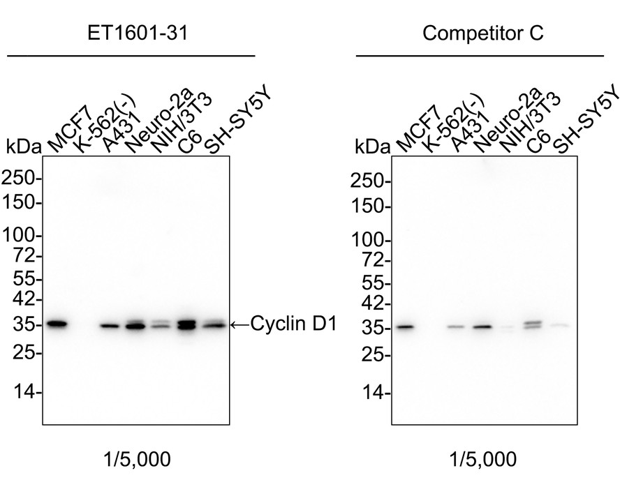
Western blot analysis of Cyclin D1 on different lysates with Rabbit anti-Cyclin D1 antibody (ET1601-31) at 1/5,000 dilution and competitor's antibody at 1/5,000 dilution.
Lane 1: MCF7 cell lysate
Lane 2: K-562 cell lysate (negative)
Lane 3: A431 cell lysate
Lane 4: Neuro-2a cell lysate
Lane 5: NIH/3T3 cell lysate
Lane 6: C6 cell lysate
Lane 7: SH-SY5Y cell lysate
Lysates/proteins at 20 µg/Lane.
Predicted band size: 34 kDa
Observed band size: 35 kDa
Exposure time: 20 seconds; ECL: K1802;
4-20% SDS-PAGE gel.
Proteins were transferred to a PVDF membrane and blocked with 5% NFDM/TBST for 1 hour at room temperature. The primary antibody (ET1601-31) at 1/5,000 dilution and competitor's antibody at 1/5,000 dilution were used in 5% NFDM/TBST at 4℃ overnight. Goat Anti-Rabbit IgG - HRP Secondary Antibody (HA1001) at 1/50,000 dilution was used for 1 hour at room temperature. -
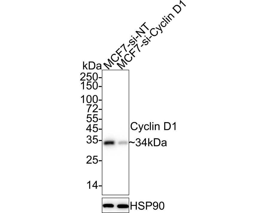
☑ Knockdown (KD)
Western blot analysis of Cyclin D1 on different lysates with Rabbit anti-Cyclin D1 antibody (ET1601-31) at 1/5,000 dilution.
Lane 1: MCF7-si NT cell lysate
Lane 2: MCF7-si Cyclin D1 cell lysate
Lysates/proteins at 10 µg/Lane.
Predicted band size: 34 kDa
Observed band size: 34 kDa
Exposure time: 17 seconds; ECL: K1801;
4-20% SDS-PAGE gel.
Proteins were transferred to a PVDF membrane and blocked with 5% NFDM/TBST for 1 hour at room temperature. The primary antibody (ET1601-31) at 1/5,000 dilution was used in 5% NFDM/TBST at 4℃ overnight. Goat Anti-Rabbit IgG - HRP Secondary Antibody (HA1001) at 1/50,000 dilution was used for 1 hour at room temperature. -
Cyclin D1 was immunoprecipitated from 0.5 mg Hela whole cell lysates with ET1601-31 at 2 μg/mL. Western blot was performed from the immunoprecipitate using ET1601-31 at 1/500 dilution for 45 minutes at room temperature. Goat anti-Rabbit IgG-HRP Secondary Antibody (HA1001) was used at 1:300,000 dilution for 30 minutes at room temperature.
Lane 1: Hela whole cell lysates at 10 μg;
Lane 2: Cyclin D1 (ET1601-31) IP in Hela whole cell lysates;
Lane 3: Rabbit IgG instead of Cyclin D1 (ET1601-31) in Hela whole cell lysates.
Predicted band size: 34 kDa
Observed band size: 34 kDa
Exposure time: 5 minutes;
12% SDS-PAGE gel. -
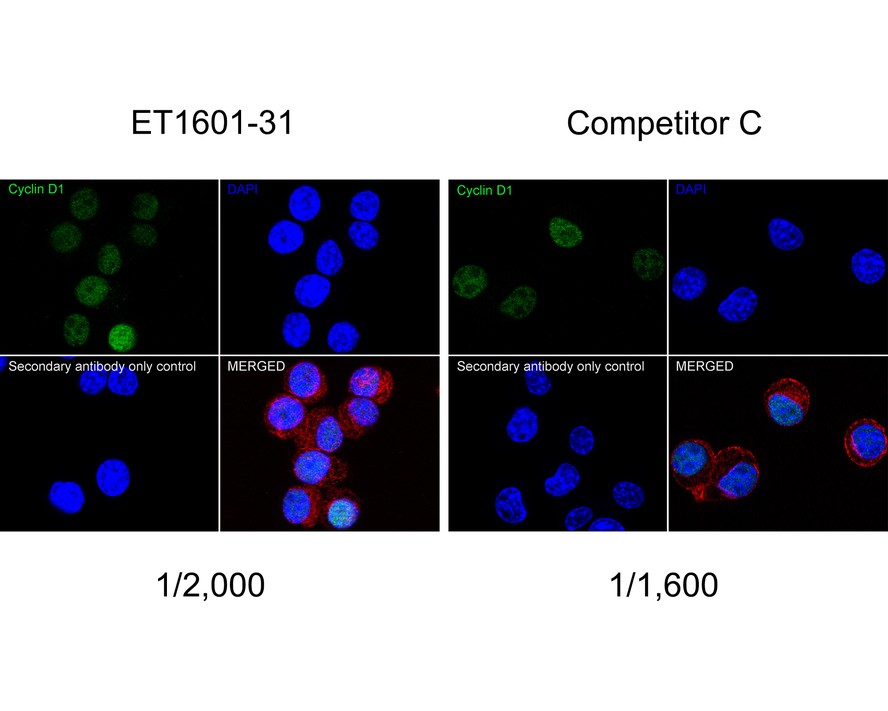
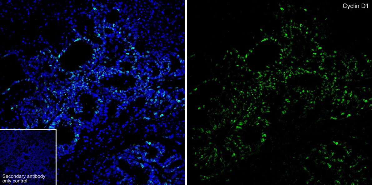
Immunocytochemistry analysis of Neuro-2a cells labeling Cyclin D1 with Rabbit anti-Cyclin D1 antibody (ET1601-31) at 1/2,000 dilution and competitor's antibody at 1/1,600 dilution.
Cells were fixed in 4% paraformaldehyde for 20 minutes at room temperature, permeabilized with 0.1% Triton X-100 in PBS for 5 minutes at room temperature, then blocked with 1% BSA in 10% negative goat serum for 1 hour at room temperature. Cells were then incubated with Rabbit anti-Cyclin D1 antibody (ET1601-31) at 1/2,000 dilution and competitor's antibody at 1/1,600 dilution in 1% BSA in PBST overnight at 4 ℃. Goat Anti-Rabbit IgG H&L (iFluor™ 488, HA1121) was used as the secondary antibody at 1/1,000 dilution. PBS instead of the primary antibody was used as the secondary antibody only control. Nuclear DNA was labelled in blue with DAPI.
Beta tubulin (M1305-2, red) was stained at 1/100 dilution overnight at +4℃. Goat Anti-Mouse IgG H&L (iFluor™ 594, HA1126) was used as the secondary antibody at 1/1,000 dilution. -
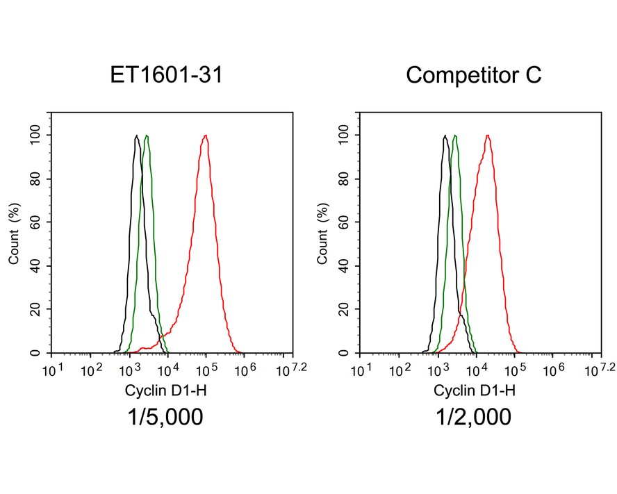
Flow cytometric analysis of MCF7 cells labeling Cyclin D1.
Cells were fixed and permeabilized. Then stained with the primary antibody (ET1601-31, red) at 1/5,000 dilution and competitor's antibody (red) at 1/2,000 dilution, compared with Rabbit IgG Isotype Control (green). After incubation of the primary antibody at +4℃ for an hour, the cells were stained with a iFluor™ 488 conjugate-Goat anti-Rabbit IgG Secondary antibody (HA1121) at 1/1,000 dilution for 30 minutes at +4℃. Unlabelled sample was used as a control (cells without incubation with primary antibody; black). -
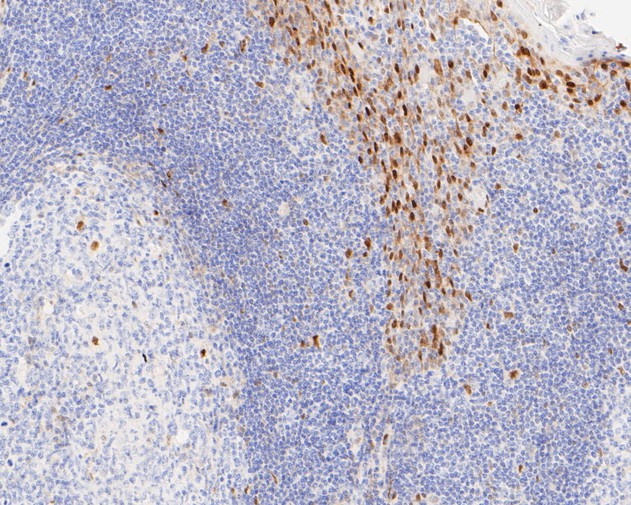
Immunohistochemical analysis of paraffin-embedded human tonsil tissue using anti-Cyclin D1 antibody. The section was pre-treated using heat mediated antigen retrieval with sodium citrate buffer (pH 6.0) for 2 minutes. The tissues were blocked in 5% BSA for 30 minutes at room temperature, washed with ddH2O and PBS, and then probed with the primary antibody (ET1601-31, 1/200) for 30 minutes at room temperature. The detection was performed using an HRP conjugated compact polymer system. DAB was used as the chromogen. Tissues were counterstained with hematoxylin and mounted with DPX.
-
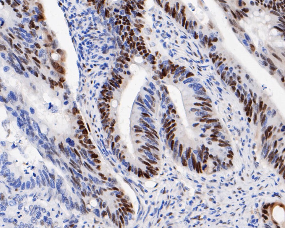
Immunohistochemical analysis of paraffin-embedded human colon carcinoma tissue using anti-Cyclin D1 antibody. The section was pre-treated using heat mediated antigen retrieval with sodium citrate buffer (pH 6.0) for 2 minutes. The tissues were blocked in 5% BSA for 30 minutes at room temperature, washed with ddH2O and PBS, and then probed with the primary antibody (ET1601-31, 1/200) for 30 minutes at room temperature. The detection was performed using an HRP conjugated compact polymer system. DAB was used as the chromogen. Tissues were counterstained with hematoxylin and mounted with DPX.
-
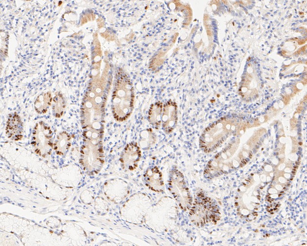
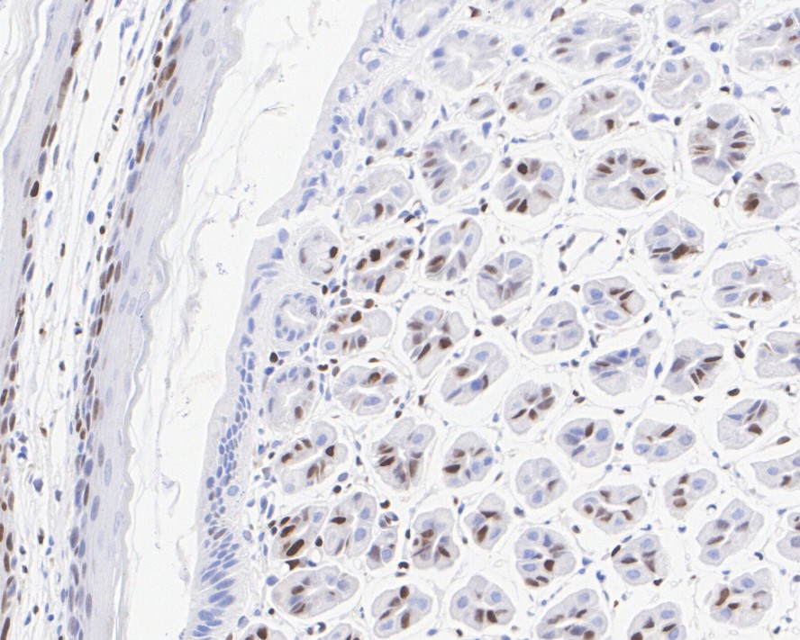
Immunohistochemical analysis of paraffin-embedded human small intestine tissue using anti-Cyclin D1 antibody. The section was pre-treated using heat mediated antigen retrieval with sodium citrate buffer (pH 6.0) for 2 minutes. The tissues were blocked in 5% BSA for 30 minutes at room temperature, washed with ddH2O and PBS, and then probed with the primary antibody (ET1601-31, 1/200) for 30 minutes at room temperature. The detection was performed using an HRP conjugated compact polymer system. DAB was used as the chromogen. Tissues were counterstained with hematoxylin and mounted with DPX.
-
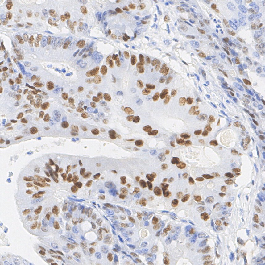
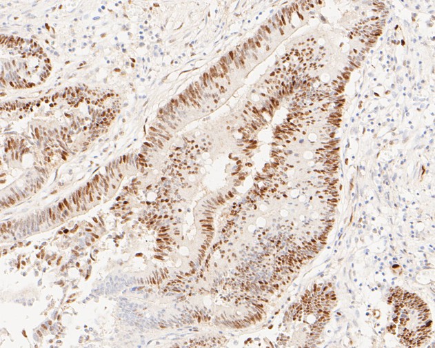
Immunohistochemical analysis of paraffin-embedded human colon carcinoma tissue using anti-Cyclin D1 antibody. The section was pre-treated using heat mediated antigen retrieval with sodium citrate buffer (pH 6.0) for 2 minutes. The tissues were blocked in 5% BSA for 30 minutes at room temperature, washed with ddH2O and PBS, and then probed with the primary antibody (ET1601-31, 1/200) for 30 minutes at room temperature. The detection was performed using an HRP conjugated compact polymer system. DAB was used as the chromogen. Tissues were counterstained with hematoxylin and mounted with DPX.
-
Immunohistochemical analysis of paraffin-embedded mouse stomach tissue with Rabbit anti-Cyclin D1 antibody (ET1601-31) at 1/1,000 dilution.
The section was pre-treated using heat mediated antigen retrieval with sodium citrate buffer (pH 6.0) for 2 minutes. The tissues were blocked in 1% BSA for 20 minutes at room temperature, washed with ddH2O and PBS, and then probed with the primary antibody (ET1601-31) at 1/1,000 dilution for 1 hour at room temperature. The detection was performed using an HRP conjugated compact polymer system. DAB was used as the chromogen. Tissues were counterstained with hematoxylin and mounted with DPX. -
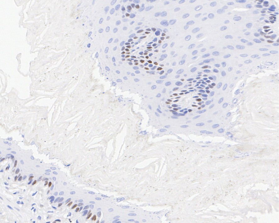
Immunohistochemical analysis of paraffin-embedded rat esophagus tissue with Rabbit anti-Cyclin D1 antibody (ET1601-31) at 1/1,000 dilution.
The section was pre-treated using heat mediated antigen retrieval with sodium citrate buffer (pH 6.0) for 2 minutes. The tissues were blocked in 1% BSA for 20 minutes at room temperature, washed with ddH2O and PBS, and then probed with the primary antibody (ET1601-31) at 1/1,000 dilution for 1 hour at room temperature. The detection was performed using an HRP conjugated compact polymer system. DAB was used as the chromogen. Tissues were counterstained with hematoxylin and mounted with DPX. -
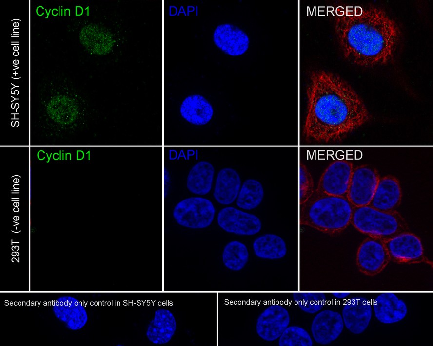
☑ Relative expression (RE)
Immunocytochemistry analysis of SH-SY5Y (positive) and 293T (negative) labeling Cyclin D1 with Rabbit anti-Cyclin D1 antibody (ET1601-31) at 1/2,000 dilution.
Cells were fixed in 4% paraformaldehyde for 20 minutes at room temperature, permeabilized with 0.1% Triton X-100 in PBS for 5 minutes at room temperature, then blocked with 1% BSA in 10% negative goat serum for 1 hour at room temperature. Cells were then incubated with Rabbit anti-Cyclin D1 antibody (ET1601-31) at 1/2,000 dilution in 1% BSA in PBST overnight at 4 ℃. Goat Anti-Rabbit IgG H&L (iFluor™ 488, HA1121) was used as the secondary antibody at 1/1,000 dilution. PBS instead of the primary antibody was used as the secondary antibody only control. Nuclear DNA was labelled in blue with DAPI.
Beta tubulin (M1305-2, red) was stained at 1/100 dilution overnight at +4℃. Goat Anti-Mouse IgG H&L (iFluor™ 594, HA1126) was used as the secondary antibody at 1/1,000 dilution. -
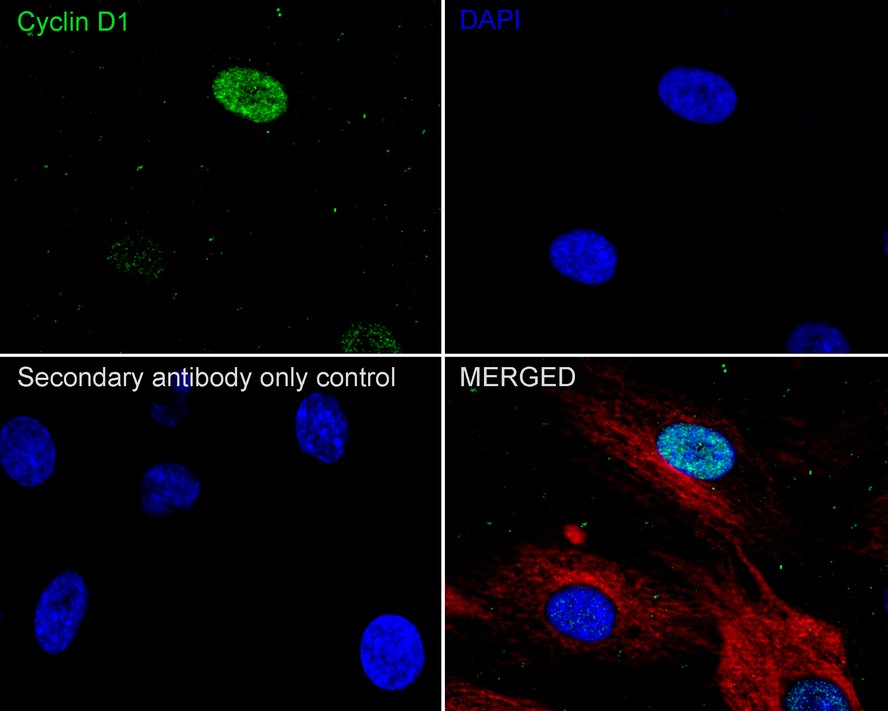
Immunocytochemistry analysis of C6 cells labeling Cyclin D1 with Rabbit anti-Cyclin D1 antibody (ET1601-31) at 1/2,000 dilution.
Cells were fixed in 4% paraformaldehyde for 20 minutes at room temperature, permeabilized with 0.1% Triton X-100 in PBS for 5 minutes at room temperature, then blocked with 1% BSA in 10% negative goat serum for 1 hour at room temperature. Cells were then incubated with Rabbit anti-Cyclin D1 antibody (ET1601-31) at 1/2,000 dilution in 1% BSA in PBST overnight at 4 ℃. Goat Anti-Rabbit IgG H&L (iFluor™ 488, HA1121) was used as the secondary antibody at 1/1,000 dilution. PBS instead of the primary antibody was used as the secondary antibody only control. Nuclear DNA was labelled in blue with DAPI.
Beta tubulin (M1305-2, red) was stained at 1/100 dilution overnight at +4℃. Goat Anti-Mouse IgG H&L (iFluor™ 594, HA1126) was used as the secondary antibody at 1/1,000 dilution. -
Cyclin D1 was immunoprecipitated from 0.2 mg MCF7 cell lysate with ET1601-31 at 2 µg/25 µl agarose. Western blot was performed from the immunoprecipitate using ET1601-31 at 1/1,000 dilution. Anti-Rabbit IgG for IP Nano-secondary antibody (NBI01H) at 1/5,000 dilution was used for 1 hour at room temperature.
Lane 1: MCF7 cell lysate (input)
Lane 2: ET1601-31 IP in MCF7 cell lysate
Lane 3: Rabbit IgG instead of ET1601-31 in MCF7 cell lysate
Blocking/Dilution buffer: 5% NFDM/TBST
Exposure time: 5 seconds; ECL: K1802
请注意: All products are "FOR RESEARCH USE ONLY AND ARE NOT INTENDED FOR DIAGNOSTIC OR THERAPEUTIC USE"
引文
-
Targeting NEDD8 in pediatric acute myeloid leukemia: an integrated bioinformatics and experimental approach
Author: Jian Sun, Cui Liu, Guangli Yang, Qian Li, Yang An, Yin Zhu, Pingping Zhang, Yaning Guan, Chang Peng, Zuochen Du, Pei Huang, Yan Chen
PMID: 40103351
期刊: Hematology
应用: WB
反应种属: Human
发表时间: 2025 Mar
-
 Citation
-
Mannose-Modified Multifunctional Iron-Based Nanozyme for Hepatocellular Carcinoma Treatment by Remodeling the Tumor Microenvironment
Author: Qi Liu, Ziwei Liang, Jiapu Wang, Yuhui Wang, Jie Wang, Shaojie Wang, Zhi Du, Liqin Zhao, Yan Wei, Di Huang
PMID: 39923382
期刊: Colloids And Surfaces B: Biointerfaces
应用: IF,WB
反应种属:
发表时间: 2025 Feb
-
Citation
-
Natural Product-Inspired Discovery of Naphthoquinone-Furo-Piperidine Derivatives as Novel STAT3 Inhibitors for the Treatment of Triple-Negative Breast Cancer
Author: Fan Chengcheng,et al
PMID: 39226127
期刊: Journal Of Medicinal Chemistry
应用: WB
反应种属: Human
发表时间: 2024 Sep
-
Citation
-
Reduced Proline-Rich Tyrosine Kinase 2 Promotes Tumor Metastasis by Activating Epithelial–Mesenchymal Transition in Colorectal Cancer
Author: Fangquan Wu ,et al
PMID: 39414740
期刊: Digestive Diseases And Sciences
应用: WB
反应种属: Human
发表时间: 2024 Oct
-
Citation
-
Hydrochlorothiazide disrupts DNA damage response to exacerbate skin photosensitivity
Author: Lei Tao,et al
PMID: 39541700
期刊: Ecotoxicology And Environmental Safety
应用: WB
反应种属: Human
发表时间: 2024 Nov
-
Citation
-
IPCEF1: Expression Patterns, Clinical Correlates and New Target of Papillary Thyroid Carcinoma
Author: Dechao Yin,et al
PMID: 39513122
期刊: Journal Of Cancer
应用: WB
反应种属: Human
发表时间: 2024 Nov
-
Citation
-
4-hydroxysesamin protects rat with right ventricular failure due to pulmonary hypertension by inhibiting JNK/p38 MAPK signaling
Author: Lingnan Zhang, Xinshun Gu
PMID: 38728253

期刊: Aging-Us
应用:
反应种属:
发表时间: 2024 May
-
Citation
-
Acetyl-11-keto-beta-boswellic acid inhibits cell proliferation and growth of oral squamous cell carcinoma via RAB7B-mediated autophagy
Author: Pan Dan,et al
PMID: 38513840
期刊: Toxicology And Applied Pharmacology
应用: WB
反应种属: Mouse
发表时间: 2024 Mar
-
Citation
-
Columbianadin suppresses glioblastoma progression by inhibiting the PI3K-Akt signaling pathway
Author: Zhang Wei,et al
PMID: 38458331
期刊: Biochemical Pharmacology
应用: WB
反应种属: Mouse
发表时间: 2024 Mar
-
Citation
-
Peptidyl-prolyl isomerase F as a prognostic biomarker associated with immune infiltrates and mitophagy in lung adenocarcinoma
Author: Zitong Feng, Lin Yuan, Luyuan Ma, Wenhao Yu, Fayez Kheir, Lukas Käsmann, Wolfgang M. Brueckl, Kai Jin, Dingxin Wang, Yi Shen, Rongyang Li, Hui Tian
PMID: 38973949

期刊: Translational Lung Cancer Research
应用:
反应种属:
发表时间: 2024 Jun
-
Citation
-
PSME3 promotes lung adenocarcinoma development by regulating the TGF-β/SMAD signaling pathway
Author: Shuai Wang, Yongmeng Li, Kai Jin, Kenichi Suda, Rongyang Li, Huiying Zhang, Hui Tian
PMID: 38973962

期刊: Translational Lung Cancer Research
应用:
反应种属:
发表时间: 2024 Jun
-
Citation
-
Traditional Medicine Xianglian Pill Suppresses high-fat diet-related colorectal cancer via inactivating TLR4/MyD88 by remodeling gut microbiota composition and bile acid metabolism
Author: Ye Chenxiao,et al
PMID: 38824980

期刊: Journal Of Ethnopharmacology
应用:
反应种属: Human
发表时间: 2024 Jun
-
Citation
-
Production of recombinant human epidermal growth factor fused with HaloTag protein and characterisation of its biological functions
Author: Bai Mengru,et al
PMID: 39035165

期刊: Peer J
应用: IF,WB
反应种属: Human
发表时间: 2024 Jul
-
Citation
-
Tenacissoside H repressed the progression of glioblastoma by inhibiting the PI3K/Akt/mTOR signaling pathway
Author: Dong Jianhong,et al
PMID: 38331340
期刊: European Journal Of Pharmacology
应用: WB
反应种属: Mouse
发表时间: 2024 Feb
-
Citation
-
BZW2 promotes malignant progression in lung adenocarcinoma through enhancing the ubiquitination and degradation of GSK3β
Author: Jin Kai,et al
PMID: 38424042

期刊: Cell Death Discovery
应用: WB
反应种属: Human
发表时间: 2024 Feb
-
Citation
-
Novel inhibitors targeting the PGK1 metabolic enzyme in glycolysis exhibit effective antitumor activity against kidney renal clear cell carcinoma in vitro and in vivo
Author: Yu He, Yinheng Luo, Lan Huang, Dan Zhang, Huijin Hou, Yue Liang, Shi Deng, Peng Zhang, Shufang Liang
PMID: 38354523

期刊: European Journal Of Medicinal Chemistry
应用: WB
反应种属: Human
发表时间: 2024 Feb
-
Citation
-
Arsenic-induced downregulation of BRWD3 suppresses proliferation and induces apoptosis in lung adenocarcinoma cells through the p53 and p65 pathways
Author: Zhu Yanhua,et al
PMID: 39190898
期刊: Human & Experimental Toxicology
应用: WB
反应种属: Human
发表时间: 2024 Aug
-
Citation
-
Integrating network pharmacology, molecular docking and experimental verification to reveal the mechanism of artesunate in inhibiting choroidal melanoma
Author: Ma Qing-yue, Liu Yi-chong, Zhang Qian, Yi Wen-dan, Sun Ying, Gao Xiao-di, Zhao Xin-tong, Wang Hao-wen, Lei Ke, Luo Wen-juan
PMID: 39185308

期刊: Frontiers In Pharmacology
应用:
反应种属:
发表时间: 2024 Aug
-
Citation
-
Sophocarpine inhibits the progression of glioblastoma via PTEN/PI3K/Akt signaling pathway
Author: Shuqiao Xing, Zhenrong Xiong, Mengmeng Wang, Yifan Li, Jiali Shi, Yiming Qian, Jia Lei, Jiamei Jia, Weiquan Zeng, Zhihui Huang, Yuanyuan Jiang
PMID: 39267674

期刊: American Journal Of Cancer Research
应用:
反应种属:
发表时间: 2024 Aug
-
Citation
-
RGS1 targeted by miR-191-3p inhibited the stemness properties of esophageal cancer cells by suppressing CXCR4/PI3K/AKT signaling
Author: Jing Xun, Yuan Ma, Botao Wang, Xiaolin Jiang, Bin Liu, Ruifang Gao, Qiongli Zhai, Runfen Cheng, Xueliang Wu, Yu Wu, Qi Zhang
PMID: 39173233

期刊: Acta Histochemica
应用: WB
反应种属: Human
发表时间: 2024 Aug
-
Citation
-
Evodiamine Inhibits the Progression of Esophageal Aquamous Cell Carcinoma via Modulating PI3K/AKT/mTOR Pathway
Author: Jiang Huangyu,et al
PMID: no pmid0505
期刊: Preprint And Has Not Been Certified By Peer Review
应用: WB
反应种属: Human,Mouse
发表时间: 2024 Apr
-
Citation
-
LTBP2 regulates cisplatin resistance in GC cells via activation of the NF-κB2/BCL3 pathway
Author: Jun Wang , Wenjia Liang , Xiangwen Wang , Zhao Chen , Lei Jiang
PMID: 38577985

期刊: Genetics And Molecular Biology
应用: WB
反应种属: Human
发表时间: 2024 Apr
-
Citation
-
3 β-Hydroxy-12-oleanen-27-oic Acid Exerts an Antiproliferative Effect on Human Colon Carcinoma HCT116 Cells via Targeting FDFT1
Author:
PMID: 37834468

期刊: International Journal Of Molecular Sciences
应用: WB
反应种属: Mouse
发表时间: 2023 Oct
-
Citation
-
ZDHHC15 promotes glioma malignancy and acts as a novel prognostic biomarker for patients with glioma
Author: Liu, Z. Y., Lan, T., Tang, F., He, Y. Z., Liu, J. S., Yang, J. Z., Chen, X., Wang, Z. F., & Li, Z. Q.
PMID: 37161425

期刊: BMC Cancer
应用: WB
反应种属: Human
发表时间: 2023 May
-
Citation
-
Angelicin impedes the progression of glioblastoma via inactivation of YAP signaling pathway
Author:
PMID: 36933380

期刊: Biomedicine & Pharmacotherapy
应用: WB
反应种属: Rat
发表时间: 2023 May
-
Citation
-
Carvedilol exhibits anti-acute T lymphoblastic leukemia effect in vitro and in vivo via inhibiting β-ARs signaling pathway
Author:
PMID: 36495764

期刊: Biochemical And Biophysical Research Communications
应用: WB
反应种属: Human
发表时间: 2023 Jan
-
Citation
-
AKIP1 accelerates glioblastoma progression through stabilizing EGFR expression
Author:
PMID: 37596322

期刊: Oncogene
应用: WB
反应种属: Mouse,Human
发表时间: 2023 Aug
-
Citation
-
SUMOylation of AnxA6 facilitates EGFR-PKCα complex formation to suppress epithelial cancer growth
Author: Zenghua Sheng, Xu Cao, Ya-nan Deng, Xinyu Zhao, Shufang Liang
PMID: 37528485

期刊: Cell Communication And Signaling
应用: WB
反应种属: Human
发表时间: 2023 Aug
-
Citation
-
TPP1 inhibits DNA damage response and chemosensitivity in esophageal cancer
Author:
PMID: 37606165

期刊: Critical Reviews In Eukaryotic Gene Expression
应用: WB
反应种属: Human
发表时间: 2023
-
Citation
-
Anti-diabetic drug canagliflozin hinders skeletal muscle regeneration in mice
Author:
PMID: 35217814

期刊: Acta Pharmacologica Sinica
应用: WB
反应种属: Mouse
发表时间: 2022 Oct
-
Citation
-
Irigenin inhibits glioblastoma progression through suppressing YAP/β-catenin signaling
Author: Xu, J., Sun, S., Zhang, W., Dong, J., Huang, C., Wang, X., Jia, M., Yang, H., Wang, Y., Jiang, Y., Cao, L., & Huang, Z.
PMID: 36532767

期刊: Frontiers In Pharmacology
应用: WB
反应种属: Human
发表时间: 2022 Nov
-
Citation
-
Cholesterol Sulfate Exerts Protective Effect on Pancreatic β-Cells by Regulating β-Cell Mass and Insulin Secretion
Author: Zhang, X., Deng, D., Cui, D., Liu, Y., He, S., Zhang, H., Xie, Y., Yu, X., Yang, S., Chen, Y., & Su, Z.
PMID: 35308228

期刊: Frontiers In Pharmacology
应用: WB
反应种属: Mouse
发表时间: 2022 Mar
-
Citation
-
ACY-1215 suppresses the proliferation and induces apoptosis of chronic myeloid leukemia cells via the ROS/PTEN/Akt pathway
Author:
PMID: 35674911

期刊: Cell Stress & Chaperones
应用: WB
反应种属: Human
发表时间: 2022 Jun
-
Citation
-
Autophagy participates in germline cyst breakdown and follicular formation by modulating glycolysis switch via Akt signaling in newly-hatched chicken ovaries
Author:
PMID: 35525303

期刊: Developmental Biology
应用: WB
反应种属: chicken
发表时间: 2022 Jul
-
Citation
-
Role of oncogene PIM-1 in the development and progression of papillary thyroid carcinoma: Involvement of oxidative stress. Molecular and cellular endocrinology, 523, 111144.
Author: Wen, Q. L., Yi, H. Q., Yang, K., Yin, C. T., Yin, W. J., Xiang, F. Y., Bao, M., Shuai, J., Song, Y. W., Ge, M. H., & Zhu, X.
PMID: 33383107

期刊: Molecular And Cellular Endocrinology
应用: WB
反应种属: Human
发表时间: 2021 Mar
-
Citation
-
Proteasome regulation by reversible tyrosine phosphorylation at the membrane
Author: Chen, L., Zhang, Y., Shu, X., Chen, Q., Wei, T., Wang, H., Wang, X., Wu, Q., Zhang, X., Liu, X., Zheng, S., Huang, L., Xiao, J., Jiang, C., Yang, B., Wang, Z., & Guo, X.
PMID: 33603165

期刊: Oncogene
应用: WB
反应种属: Human
发表时间: 2021 Mar
-
Citation
-
A MYBL2 complex for RRM2 transactivation and the synthetic effect of MYBL2 knockdown with WEE1 inhibition against colorectal cancer. Cell death & disease, 12(7), 683.
Author: Liu, Q., Guo, L., Qi, H., Lou, M., Wang, R., Hai, B., Xu, K., Zhu, L., Ding, Y., Li, C., Xie, L., Shen, J., Xiang, X., & Shao, J.
PMID: 34234118

期刊: Cell Death & Disease
应用: WB
反应种属: Mouse
发表时间: 2021 Jul
-
Citation
-
All-Trans Retinoic Acid Potentiates Antitumor Efficacy of Cisplatin by Increasing Differentiation of Cancer Stem-Like Cells in Cervical Cancer. Annals of clinical and laboratory science, 51(1), 22–29.
Author: Fan, W. J., Ding, H., Chen, X. X., & Yang, L.
PMID: 33653777
期刊: Annals Of Clinical And Laboratory Science
应用:
反应种属:
发表时间: 2021 Jan
-
Citation
-
Inhibition of cyclooxygenase-2 enhanced intestinal epithelial homeostasis via suppressing β-catenin signalling pathway in experimental liver fibrosis
Author:
PMID: 34145945

期刊: Journal Of Cellular And Molecular Medicine
应用: WB,IHC
反应种属: Mouse
发表时间: 2021 Aug
-
Citation
-
Canagliflozin impairs blood reperfusion of ischaemic lower limb partially by inhibiting the retention and paracrine function of bone marrow derived mesenchymal stem cells
Author: Zhengzheng Li;Zhiting Wang
PMID: 31981975
期刊: eBioMedicine
应用: WB
反应种属: Mouse
发表时间: 2020 Feb
-
Citation
-
Tex10 promotes stemness and EMT phenotypes in esophageal squamous cell carcinoma via the Wnt/β-catenin pathway
Author: Gang Feng
PMID: 31638260
期刊: Oncology Reports
应用: WB
反应种属: Human
发表时间: 2019 Dec
-
Citation
同靶点 & 同通路的产品

Cyclin D1 Rabbit Polyclonal Antibody
Application: WB,IF-Cell,FC
Reactivity: Human,Mouse,Rat,Zebrafish
Conjugate: unconjugated

Cyclin D1 Rabbit Polyclonal Antibody
Application: WB,IHC-P,FC
Reactivity: Human,Mouse,Rat
Conjugate: unconjugated
