CD161 Mouse Monoclonal Antibody [A8E1]

Catalog# HA601063
CD161 Mouse Monoclonal Antibody [A8E1]
-
WB
-
IHC-P
-
FC
-
Human
概述
产品名称
CD161 Mouse Monoclonal Antibody [A8E1]
抗体类型
Mouse Monoclonal Antibody
免疫原
Synthetic peptide within human CD161 aa 176-225.
种属反应性
Human
验证应用
WB, IHC-P, FC
分子量
Predicted band size: 25 kDa
阳性对照
THP-1 cell lysate, HL-60 cell lysate, K-562 cell lysate, Jurkat cell lysate, human kidney tissue lysate, human kidney tissue, THP-1.
偶联
unconjugated
克隆号
A8E1
RRID
产品特性
形态
Liquid
浓度
1.1ug/ul
存放说明
Store at +4℃ after thawing. Aliquot store at -20℃. Avoid repeated freeze / thaw cycles.
存储缓冲液
PBS (pH7.4), 0.1% BSA, 40% Glycerol. Preservative: 0.05% Sodium Azide.
亚型
IgG2b
纯化方式
Protein A affinity purified.
应用稀释度
-
WB
-
1:2,000
-
IHC-P
-
1:500
-
FC
-
1:500-1:1,000
靶点
功能
Killer cell lectin-like receptor subfamily B, member 1, also known as KLRB1, NKR-P1A or CD161 (cluster of differentiation 161), is a human gene. Natural killer (NK) cells are lymphocytes that mediate cytotoxicity and secrete cytokines after immune stimulation. Several genes of the C-type lectin superfamily, including the rodent NKRP1 family of glycoproteins, are expressed by NK cells and may be involved in the regulation of NK cell function. The KLRB1 protein contains an extracellular domain with several motifs characteristic of C-type lectins, a transmembrane domain, and a cytoplasmic domain. The KLRB1 protein, NKR-P1A or CD161, is classified as a type II membrane protein because it has an external C terminus. NKR-P1A, the receptor encoded by the KLRB1 gene, recognizes Lectin Like Transcript-1 (LLT1) as a functional ligand. Expressed in a subset of NK cells predominantly in intestinal epithelium and liver. Detected in peripheral blood T-cells and preferentially in adult T-cells with a memory antigenic phenotype.
背景文献
1. Christiansen D., Mouhtouris E., Milland J., Zingoni A., Santoni A., Sandrin M.S. Recognition of a carbohydrate xenoepitope by human NKRP1A (CD161). Xenotransplantation 13:440-446(2006).
2. Pozo D., Vales-Gomez M., Mavaddat N., Williamson S.C., Chisholm S.E., Reyburn H. CD161 (human NKR-P1A) signaling in NK cells involves the activation of acid sphingomyelinase. J. Immunol. 176:2397-2406(2006).
亚细胞定位
Membrane
UNIPROT #
别名
C-type lectin domain family 5 member B antibody
CD161 antibody
CLEC5B antibody
HNKR-P1a antibody
Killer Cell Lectin like Receptor Subfamily B Member 1 antibody
Killer cell lectin-like receptor subfamily B member 1 antibody
KLRB1 antibody
KLRB1_HUMAN antibody
Natural killer cell surface protein P1A antibody
NKR antibody
展开图片
-
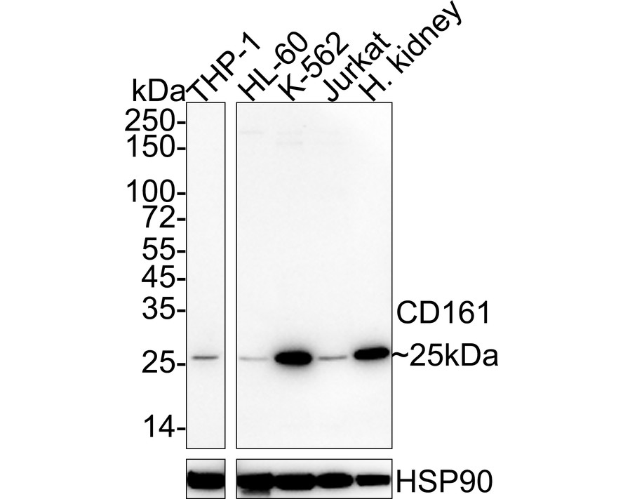
Western blot analysis of CD161 on different lysates with Mouse anti-CD161 antibody (HA601063) at 1/2,000 dilution.
Lane 1: THP-1 cell lysate (20 µg/Lane)
Lane 2: HL-60 cell lysate (20 µg/Lane)
Lane 3: K-562 cell lysate (20 µg/Lane)
Lane 4: Jurkat cell lysate (20 µg/Lane)
Lane 5: Human kidney tissue lysate (40 µg/Lane)
Predicted band size: 25 kDa
Observed band size: 25 kDa
Exposure time: 3 minutes; ECL: K1801;
4-20% SDS-PAGE gel.
Proteins were transferred to a PVDF membrane and blocked with 5% NFDM/TBST for 1 hour at room temperature. The primary antibody (HA601063) at 1/2,000 dilution was used in 5% NFDM/TBST at 4℃ overnight. Anti-Mouse IgG for IP Nano-secondary antibody (NBI02H) at 1/5,000 dilution was used for 1 hour at room temperature. -
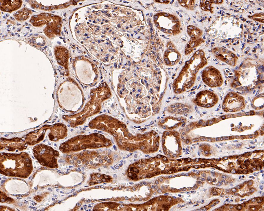
Immunohistochemical analysis of paraffin-embedded human kidney tissue with Mouse anti-CD161 antibody (HA601063) at 1/500 dilution.
The section was pre-treated using heat mediated antigen retrieval with Tris-EDTA buffer (pH 9.0) for 20 minutes. The tissues were blocked in 1% BSA for 20 minutes at room temperature, washed with ddH2O and PBS, and then probed with the primary antibody (HA601063) at 1/500 dilution for 1 hour at room temperature. The detection was performed using an HRP conjugated compact polymer system. DAB was used as the chromogen. Tissues were counterstained with hematoxylin and mounted with DPX. -
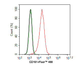
Flow cytometric analysis of THP-1 cells labeling CD161.
Cells were washed twice with cold PBS and resuspend. Then stained with the primary antibody (HA601063, 1ug/ml) (red) compared with Mouse IgG1 Isotype Control (green). After incubation of the primary antibody at +4℃ for 30 minutes, the cells were stained with a iFluor™ 488 conjugate-Goat anti-Mouse IgG Secondary antibody (HA1125) at 1/1,000 dilution for 30 minutes at +4℃. Unlabelled sample was used as a control (cells without incubation with primary antibody; black).
Please note: All products are "FOR RESEARCH USE ONLY AND ARE NOT INTENDED FOR DIAGNOSTIC OR THERAPEUTIC USE"