兔多克隆抗体的特点与产品推荐
抗体是一种由免疫系统产生的蛋白质分子,也被称为免疫球蛋白。它们由免疫细胞中的B淋巴细胞合成,在免疫应答中起到重要作用。抗原是指能够激发免疫系统产生抗体或T细胞应答的物质,可以是细菌、病毒、真菌、寄生虫等微生物,也可以是体内异常细胞或外来物质。每种抗体通常只能与特定的抗原结合。抗体根据结构和功能的不同可以被分为五大类:一、IgG,最常见的抗体类型,可在体内提供长期保护;二、IgM,在第一次免疫应答时产生,能够快速识别并结合抗原;三、IgA,主要存在于黏膜表面,提供局部的免疫保护;四、IgE,通常参与过敏反应;五、IgD,具体功能仍在研究中,目前的研究表明可能参与免疫体系调节。
什么是兔多克隆抗体?
兔多克隆抗体是一种通过对兔子免疫所产生的多克隆抗体。多克隆抗体是指在免疫动物体内通过免疫原刺激,由多个B细胞克隆产生的抗体群体。兔子因其免疫系统对抗原具有较强的免疫应答成为最常用的动物模型之一,它在接种抗原后能快速产生高亲和力的多克隆抗体。目前,兔多克隆抗体在科研、诊断等领域广泛应用,常用于免疫组织化学、免疫印迹、免疫荧光等实验技术中,用于检测和定位特定的抗原。
兔多克隆抗体的生产
兔多克隆抗体的生产体系已相对完善,通常分为以下五个步骤:
一、免疫原准备。确定需要制备抗体的抗原,抗原可以是蛋白质、多肽、多糖或其他分子。
二、兔子免疫。将抗原注射到兔子体内,通常通过皮下、肌肉或静脉注射。同时为了增强免疫效果,常会使用佐剂与抗原混合注射。
三、免疫程序。需要将抗原间隔一定时间多次注射,使兔子免疫应答产生足够的抗体。
四、血清采集、分离与纯化。等待兔子产生足够的抗体后,通过耳部或静脉采集血液样本。将血液样本离心分离后收集血清,再通过纯化去除其他无关物质获得抗体。常用的纯化方式包括蛋白A/G亲和纯化,离子交换层析、凝胶过滤等。
五、抗体的保存。纯化后的抗体通常与抗保混合,-20℃或-80℃冷冻保存以延长其稳定性和使用时限。
兔多克隆抗体的特点
一、多样性。兔子的免疫系统对抗原有很强的免疫应答能力,能够产生多个不同的抗体克隆。这表示着每个抗体克隆能够识别抗原的不同表位,提高了抗体的多样性。
二、周期短、成本低。相对于其他动物模型,兔子可以更快产生免疫应答。这意味着在制备时,可以较快的获得抗体,节省时间;此外,兔子相对小鼠等小型动物模型而言,体型较大,产生的抗体量也相对较高。这意味着从每次免疫中可以获得较多的抗体,用于商业化生产成本较低。
三、高亲和力。兔多克隆抗体的基因片段相对较长,往往具有很高的亲和力,能够更好地结合目标抗原,提高检测或治疗的敏感性和效果。
四、兔多克隆抗体优势显著,但缺点也不容忽视。首先,不同批次间不可避免地存在差异,当其用于商业化抗体生产中则会产生供货限制问题,无法保证每个批次间具有相同的灵敏度。其次,兔多克隆抗体在免疫过程中易产生非特异性抗体,这些抗体在应用中会产生一定的干扰。
四、兔多克隆抗体优势显著,但缺点也不容忽视。首先,不同批次间不可避免地存在差异,当其用于商业化抗体生产中则会产生供货限制问题,无法保证每个批次间具有相同的灵敏度。其次,兔多克隆抗体在免疫过程中易产生非特异性抗体,这些抗体在应用中会产生一定的干扰。
华安兔多抗产品推荐

Lane 1: HeLa cell lysate
Lane 2: MCF7 cell lysate
Lane 3: HEK-293 cell lysate
Lysates/proteins at 15 µg/Lane.
Predicted band size: 21 kDa
Observed band size: 21 kDa
Exposure time: 1 minute 34 seconds;
4-20% SDS-PAGE gel.
Proteins were transferred to a PVDF membrane and blocked with 5% NFDM/TBST for 1 hour at room temperature. The primary antibody (ER0907) at 1/5,000 dilution and competitor's antibody at 1/1,000 dilution were used in 5% NFDM/TBST at 4℃ overnight. Goat Anti-Rabbit IgG - HRP Secondary Antibody (HA1001) at 1/50,000 dilution was used for 1 hour at room temperature.
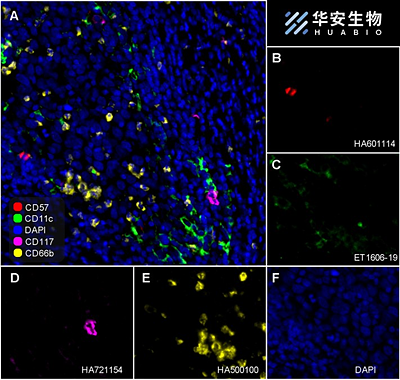
Fluorescence multiplex immunohistochemical analysis of the human cervical cancer (Formalin/PFA-fixed paraffin-embedded sections). Panel A: the merged image of anti-CD57 (HA601114, red), anti-CD11c (ET1606-19, green), anti-CD117 (HA21154, magenta) and anti-CD66b (HA500100, yellow) on human cervical cancer. Panel B: anti- CD57 stained on NKT cells. Panel C: anti-CD11c stained on dendritic cells. Panel D: anti-CD117 stained on mast cells. Panel E: anti-CD66b stained on neutrophils. HRP Conjugated UltraPolymer Goat Polyclonal Antibody HA1119/HA1120 was used as a secondary antibody. The immunostaining was performed with the Sequential Immuno-staining Kit (IRISKit™MH010101, www.luminiris.cn). The section was incubated in four rounds of staining: in the order of HA601114 (1/500 dilution), ET1606-19 (1/1,000 dilution), HA721154 (1/1,000 dilution), and HA500100 (1/1,000 dilution) for 20 mins at room temperature. Each round was followed by a separate fluorescent tyramide signal amplification system. Heat mediated antigen retrieval with Tris-EDTA buffer (pH 9.0) for 30 mins at 95C. DAPI (blue) was used as a nuclear counter stain. Image acquisition was performed with Olympus VS200 Slide Scanner.
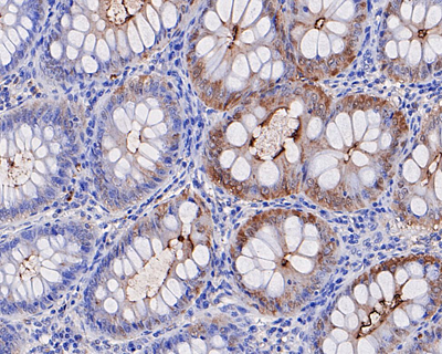
Immunohistochemical analysis of paraffin-embedded human colon tissue using anti-CD66b antibody. The section was pre-treated using heat mediated antigen retrieval with sodium citrate buffer (pH 6.0) for 20 minutes. The tissues were blocked in 5% BSA for 30 minutes at room temperature, washed with ddH2O and PBS, and then probed with the primary antibody (HA500100, 1/400) for 30 minutes at room temperature. The detection was performed using an HRP conjugated compact polymer system. DAB was used as the chromogen. Tissues were counterstained with hematoxylin and mounted with DPX.