Progesterone Receptor Recombinant Rabbit Monoclonal Antibody [JF0549]


Catalog# ET1702-24
Progesterone Receptor Recombinant Rabbit Monoclonal Antibody [JF0549]
-
WB
-
IF-Cell
-
IF-Tissue
-
IHC-P
-
IP
-
Human
概述
产品名称
Progesterone Receptor Recombinant Rabbit Monoclonal Antibody [JF0549]
抗体类型
Recombinant Rabbit monoclonal Antibody
免疫原
Synthetic peptide within N-terminal human Progesterone Receptor.
种属反应性
Human
验证应用
WB, IF-Cell, IF-Tissue, IHC-P, IP
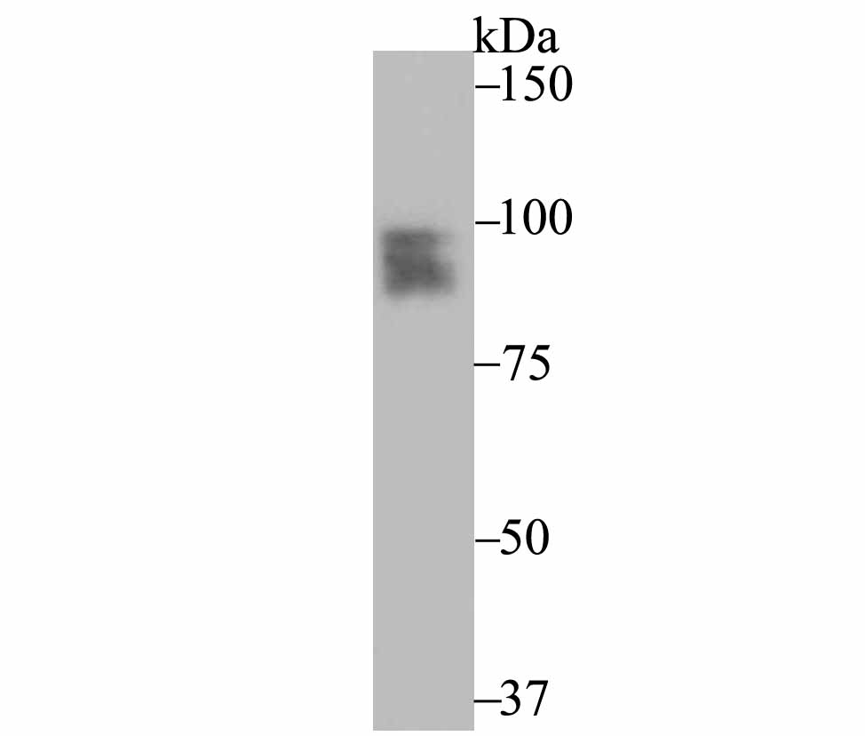
分子量
99 kDa
阳性对照
MCF-7, human breast tissue, human cervix tissue, human uterus tissue, human smooth muscle tissue.
偶联
unconjugated
克隆号
JF0549
RRID
产品特性
形态
Liquid
浓度
1ug/ul
存放说明
Store at +4℃ after thawing. Aliquot store at -20℃ or -80℃. Avoid repeated freeze / thaw cycles.
存储缓冲液
1*TBS (pH7.4), 0.05% BSA, 40% Glycerol. Preservative: 0.05% Sodium Azide.
亚型
IgG
纯化方式
Protein A affinity purified.
应用稀释度
-
WB
-
1:1,000
-
IF-Cell
-
1:50-1:100
-
IF-Tissue
-
1:50-1:100
-
IHC-P
-
1:50-1:200
-
IP
-
Use at an assay dependent concentration.
靶点
功能
PR, a protein with 946 amino acids, is a ligand-activated transcription factor member of the steroid receptor super family of nuclear receptors. The functional structure is similar to that of estrogen receptor (ER), with considerable sequence homology in the DNA-binding central domain. PR is predominantly expressed in tumours of female sex steroid responsive tissues such as the mammary gland, endometrium and the ovary. About half of the breast carcinomas are ER+/PR+. A small fraction (<5%) is ER-/PR+. About half of the non-mucinous ovarian carcinomas are also PR+. From other PR-expressing tumours, meningiomas, various pancreatic neoplasms such as solid-pseudopapillary tumour and endocrine tumours, and salivary gland neoplasms are worth mentioning. The ER and PR status has been used for over 20 years as a predictor of breast carcinoma responsiveness to endocrine therapy and as a prognostic indicator for early recurrence. Up to 75% of ER+/PR+ breast carcinomas respond positively to endocrine treatment. ER+/PR- tumours are less responsive, and thus PR status adds information to ER-status. In combination the two predict benefit from endocrine therapy both in adjuvant setting and in advanced disease. In breast cancer predominance of one isoform, namely PR-B, is common. The majority of endometrial carcinomas express only one isoform. The applications of antibodies to PR are similar to those against ER, i.e. diagnosis of PR-positive tumours (often metastasis) and prediction of therapeutic response of breast carcinoma.
背景文献
1. Bondi CD et al. The effect of estradiol, progesterone, and melatonin on estrous cycling and ovarian aromatase expression in intact female mice. Eur J Obstet Gynecol Reprod Biol 174:80-5 (2014).
2. Yu Y et al. Prostate stromal cells express the progesterone receptor to control cancer cell mobility. PLoS One 9:e92714 (2014).
序列相似性
Belongs to the nuclear hormone receptor family. NR3 subfamily.
组织特异性
In reproductive tissues the expression of isoform A and isoform B varies as a consequence of developmental and hormonal status. Isoform A and isoform B are expressed in comparable levels in uterine glandular epithelium during the proliferative phase of the menstrual cycle. Expression of isoform B but not of isoform A persists in the glands during mid-secretory phase. In the stroma, isoform A is the predominant form throughout the cycle. Heterogeneous isoform expression between the glands of the endometrium basalis and functionalis is implying region-specific responses to hormonal stimuli.
翻译后修饰
Phosphorylated on multiple serine sites. Several of these sites are hormone-dependent. Phosphorylation on Ser-294 occurs preferentially on isoform B, is highly hormone-dependent and modulates ubiquitination and sumoylation on Lys-388. Phosphorylation on Ser-102 and Ser-345 also requires induction by hormone. Basal phosphorylation on Ser-81, Ser-162, Ser-190 and Ser-400 is increased in response to progesterone and can be phosphorylated in vitro by the CDK2-A1 complex. Increased levels of phosphorylation on Ser-400 also in the presence of EGF, heregulin, IGF, PMA and FBS. Phosphorylation at this site by CDK2 is ligand-independent, and increases nuclear translocation and transcriptional activity. Phosphorylation at Ser-162 and Ser-294, but not at Ser-190, is impaired during the G(2)/M phase of the cell cycle. Phosphorylation on Ser-345 by ERK1/2 MAPK is required for interaction with SP1.; Sumoylation is hormone-dependent and represses transcriptional activity. Sumoylation on all three sites is enhanced by PIAS3. Desumoylated by SENP1. Sumoylation on Lys-388, the main site of sumoylation, is repressed by ubiquitination on the same site, and modulated by phosphorylation at Ser-294.; Ubiquitination is hormone-dependent and represses sumoylation on the same site. Promoted by MAPK-mediated phosphorylation on Ser-294.; Palmitoylated by ZDHHC7 and ZDHHC21. Palmitoylation is required for plasma membrane targeting and for rapid intracellular signaling via ERK and AKT kinases and cAMP generation.
亚细胞定位
Nucleus, Cytoplasm, Mitochondrion outer membrane.
UNIPROT #
别名
NR3C3 antibody
Nuclear receptor subfamily 3 group C member 3 antibody
PGR antibody
PR antibody
PRA antibody
PRB antibody
PRGR_HUMAN antibody
Progesterone receptor antibody
Progestin receptor form A antibody
Progestin receptor form B antibody
图片
-
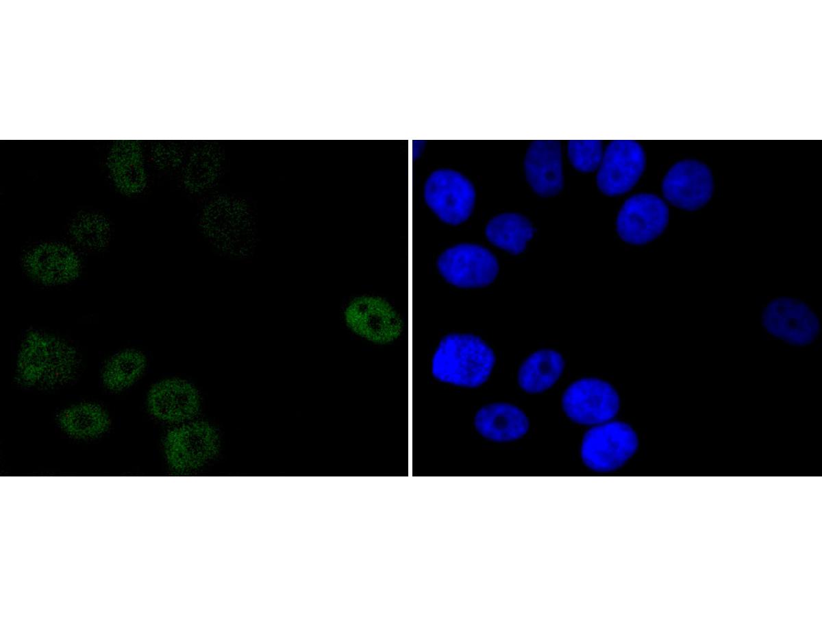
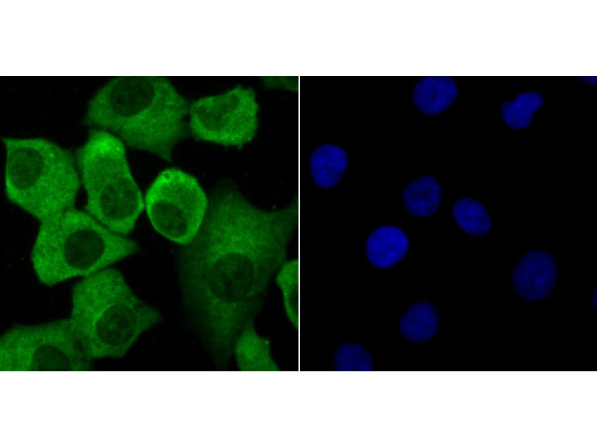
ICC staining of Progesterone Receptor in MCF-7 cells (green). Formalin fixed cells were permeabilized with 0.1% Triton X-100 in TBS for 10 minutes at room temperature and blocked with 1% Blocker BSA for 15 minutes at room temperature. Cells were probed with the primary antibody (ET1702-24, 1/50) for 1 hour at room temperature, washed with PBS. Alexa Fluor®488 Goat anti-Rabbit IgG was used as the secondary antibody at 1/1,000 dilution. The nuclear counter stain is DAPI (blue).
-
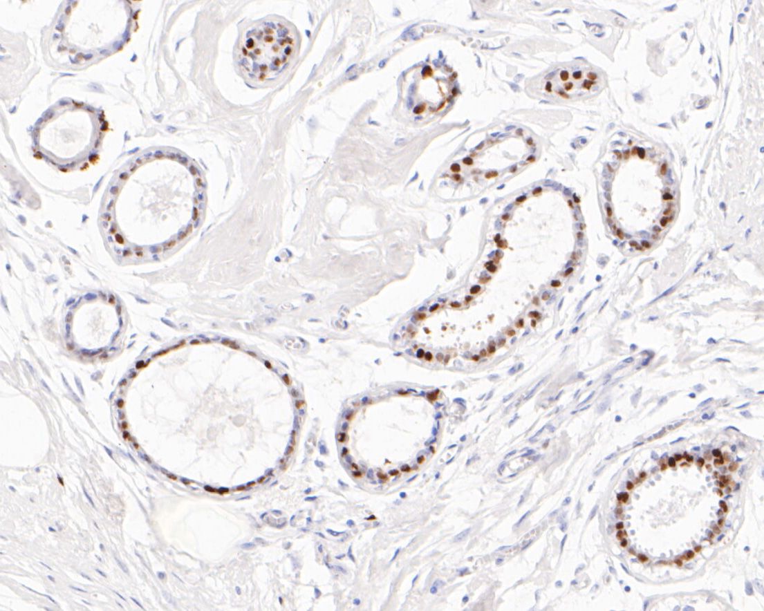
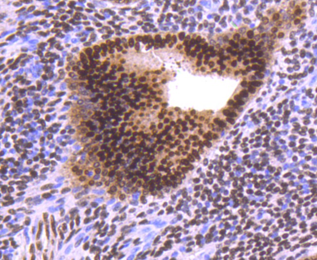
Immunohistochemical analysis of paraffin-embedded human breast tissue using anti-Progesterone Receptor antibody. The section was pre-treated using heat mediated antigen retrieval with sodium citrate buffer (pH 6.0) for 20 minutes. The tissues were blocked in 5% BSA for 30 minutes at room temperature, washed with ddH2O and PBS, and then probed with the primary antibody (ET1702-24, 1/50) for 30 minutes at room temperature. The detection was performed using an HRP conjugated compact polymer system. DAB was used as the chromogen. Tissues were counterstained with hematoxylin and mounted with DPX.
-
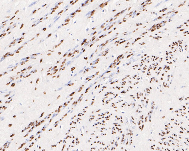
Immunohistochemical analysis of paraffin-embedded human cervix tissue using anti-Progesterone Receptor antibody. The section was pre-treated using heat mediated antigen retrieval with sodium citrate buffer (pH 6.0) for 20 minutes. The tissues were blocked in 5% BSA for 30 minutes at room temperature, washed with ddH2O and PBS, and then probed with the primary antibody (ET1702-24, 1/50) for 30 minutes at room temperature. The detection was performed using an HRP conjugated compact polymer system. DAB was used as the chromogen. Tissues were counterstained with hematoxylin and mounted with DPX.
-
Immunohistochemical analysis of paraffin-embedded human uterus tissue using anti-Progesterone Receptor antibody. The section was pre-treated using heat mediated antigen retrieval with sodium citrate buffer (pH 6.0) for 20 minutes. The tissues were blocked in 5% BSA for 30 minutes at room temperature, washed with ddH2O and PBS, and then probed with the primary antibody (ET1702-24, 1/50) for 30 minutes at room temperature. The detection was performed using an HRP conjugated compact polymer system. DAB was used as the chromogen. Tissues were counterstained with hematoxylin and mounted with DPX.
-
Immunohistochemical analysis of paraffin-embedded human breast tissue using anti-Progesterone Receptor antibody. The section was pre-treated using heat mediated antigen retrieval with sodium citrate buffer (pH 6.0) for 20 minutes. The tissues were blocked in 5% BSA for 30 minutes at room temperature, washed with ddH2O and PBS, and then probed with the primary antibody (ET1702-24, 1/100) for 30 minutes at room temperature. The detection was performed using an HRP conjugated compact polymer system. DAB was used as the chromogen. Tissues were counterstained with hematoxylin and mounted with DPX.
-
Immunohistochemical analysis of paraffin-embedded human smooth muscle tissue using anti-Progesterone Receptor antibody. The section was pre-treated using heat mediated antigen retrieval with sodium citrate buffer (pH 6.0) for 20 minutes. The tissues were blocked in 5% BSA for 30 minutes at room temperature, washed with ddH2O and PBS, and then probed with the primary antibody (ET1702-24, 1/100) for 30 minutes at room temperature. The detection was performed using an HRP conjugated compact polymer system. DAB was used as the chromogen. Tissues were counterstained with hematoxylin and mounted with DPX.
Please note: All products are "FOR RESEARCH USE ONLY AND ARE NOT INTENDED FOR DIAGNOSTIC OR THERAPEUTIC USE"
同靶点&同通路的产品
Progesterone Receptor Rabbit Polyclonal Antibody
Application: IF-Cell,IHC-P
Reactivity: Human,Mouse
Conjugate: unconjugated

Progesterone receptor Rabbit Polyclonal Antibody
Application: WB,IF-Cell,FC
Reactivity: Human
Conjugate: unconjugated