PAX5 Recombinant Rabbit Monoclonal Antibody [JJ08-87]


Catalog# ET1701-49
PAX5 Recombinant Rabbit Monoclonal Antibody [JJ08-87]
-
WB
-
IF-Cell
-
IF-Tissue
-
IHC-P
-
FC
-
Human
-
Mouse
-
Rat
概述
产品名称
PAX5 Recombinant Rabbit Monoclonal Antibody [JJ08-87]
抗体类型
Recombinant Rabbit monoclonal Antibody
免疫原
Synthetic peptide within human PAX5 aa 240-280.
种属反应性
Human, Mouse, Rat
验证应用
WB, IF-Cell, IF-Tissue, IHC-P, FC
分子量
Predicted band size: 42 kDa
阳性对照
Ramos cell lysate, Raji cell lysate, Daudi cell lysates, Hela, MCF-7, HepG2, human lymph nodes tissue, human tonsil tissue, human spleen tissue, mouse spleen tissue, rat spleen tissue, Raji.
偶联
unconjugated
克隆号
JJ08-87
RRID
产品特性
形态
Liquid
浓度
1ug/ul
存放说明
Store at +4℃ after thawing. Aliquot store at -20℃ or -80℃. Avoid repeated freeze / thaw cycles.
存储缓冲液
1*TBS (pH7.4), 0.05% BSA, 40% Glycerol. Preservative: 0.05% Sodium Azide.
亚型
IgG
纯化方式
Protein A affinity purified.
应用稀释度
-
WB
-
1:5,000
-
IF-Cell
-
1:50-1:200
-
IF-Tissue
-
1:50-1:200
-
IHC-P
-
1:400-1:1,000
-
FC
-
1:50-1:100
靶点
功能
The Pax family of nuclear transcription factors is comprised of nine members that function during embryogenesis to regulate the temporal and position-dependent differentiation of cells. Pax family genes are also involved in a variety of signal transduction pathways in the adult organism. Mutations in Pax proteins have been linked to disease and cancer in humans. For example, the human PAX5 gene encodes a B cell lineage-specific protein, Pax-5, also designated B cell specific activator protein or BSAP, which is expressed in pro-B, pre-B and mature B lymphocytes but not in plasma cells. Pax-5 functions to regulate not only B cell development, but also influences the balance between immunoglobulin secretion and B cell proliferation. Overexpression of Pax-5 has been implicated in cellular transformation, and in the case of small lymphocytic lymphomas with plasmacytoid differentiation, a t(9;14)(p13;q32) translocation resulting in the deregulation of PAX5 gene expression has been detected.
背景文献
1. Zhao L et al. Paired box 5 is a frequently methylated lung cancer tumour suppressor gene interfering -catenin signalling and GADD45G expression. J Cell Mol Med 20:842-54 (2016).
2. Ren Y et al. Diagnostic utility of PAX2 and PAX5 in distinguishing non-small cell lung cancer from small cell lung cancer. Int J Clin Exp Pathol 8:14709-16 (2015).
翻译后修饰
O-glycosylated.
亚细胞定位
Nucleus.
UNIPROT #
别名
B cell lineage specific activator antibody
B cell lineage specific activator protein antibody
B cell specific activator protein antibody
B cell specific transcription factor antibody
B-cell-specific transcription factor antibody
BSAP antibody
EBB-1 antibody
KLP antibody
Paired box 5 antibody
Paired box gene 5 (B cell lineage specific activator protein) antibody
展开图片
-
Western blot analysis of PAX5 on different lysates with Rabbit anti-PAX5 antibody (ET1701-49) at 1/5,000 dilution.
Lane 1: Ramos cell lysate
Lane 2: Raji cell lysate
Lane 3: Daudi cell lysate
Lysates/proteins at 20 µg/Lane.
Predicted band size: 42 kDa
Observed band size: 50 kDa
Exposure time: 3 minutes 10 seconds;
4-20% SDS-PAGE gel.
Proteins were transferred to a PVDF membrane and blocked with 5% NFDM/TBST for 1 hour at room temperature. The primary antibody (ET1701-49) at 1/5,000 dilution was used in 5% NFDM/TBST at 4℃ overnight. Goat Anti-Rabbit IgG - HRP Secondary Antibody (HA1001) at 1:50,000 dilution was used for 1 hour at room temperature. -
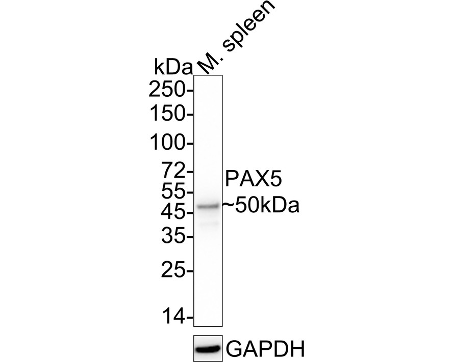
Western blot analysis of PAX5 on Mouse spleen tissue lysates with Rabbit anti-PAX5 antibody (ET1701-49) at 1/5,000 dilution.
Lysates/proteins at 40 µg/Lane.
Predicted band size: 42 kDa
Observed band size: 50 kDa
Exposure time: 30 seconds; ECL: K1801;
4-20% SDS-PAGE gel.
Proteins were transferred to a PVDF membrane and blocked with 5% NFDM/TBST for 1 hour at room temperature. The primary antibody (ET1701-49) at 1/5,000 dilution was used in 5% NFDM/TBST at 4℃ overnight. Goat Anti-Rabbit IgG - HRP Secondary Antibody (HA1001) at 1/50,000 dilution was used for 1 hour at room temperature. -
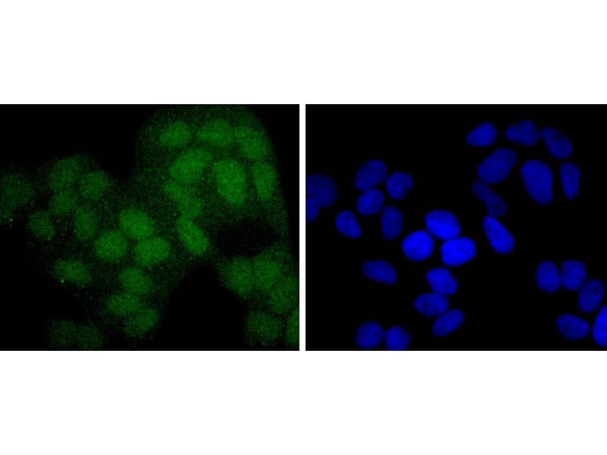
ICC staining of PAX5 in Hela cells (green). Formalin fixed cells were permeabilized with 0.1% Triton X-100 in TBS for 10 minutes at room temperature and blocked with 10% negative goat serum for 15 minutes at room temperature. Cells were probed with the primary antibody (ET1701-49, 1/50) for 1 hour at room temperature, washed with PBS. Alexa Fluor®488 conjugate-Goat anti-Rabbit IgG was used as the secondary antibody at 1/1,000 dilution. The nuclear counter stain is DAPI (blue).
-
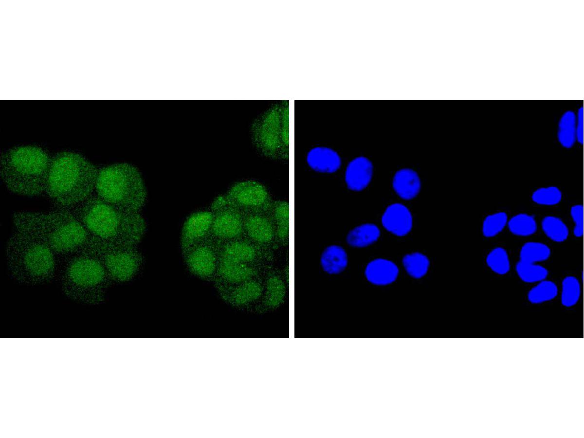
ICC staining of PAX5 in MCF-7 cells (green). Formalin fixed cells were permeabilized with 0.1% Triton X-100 in TBS for 10 minutes at room temperature and blocked with 10% negative goat serum for 15 minutes at room temperature. Cells were probed with the primary antibody (ET1701-49, 1/50) for 1 hour at room temperature, washed with PBS. Alexa Fluor®488 conjugate-Goat anti-Rabbit IgG was used as the secondary antibody at 1/1,000 dilution. The nuclear counter stain is DAPI (blue).
-
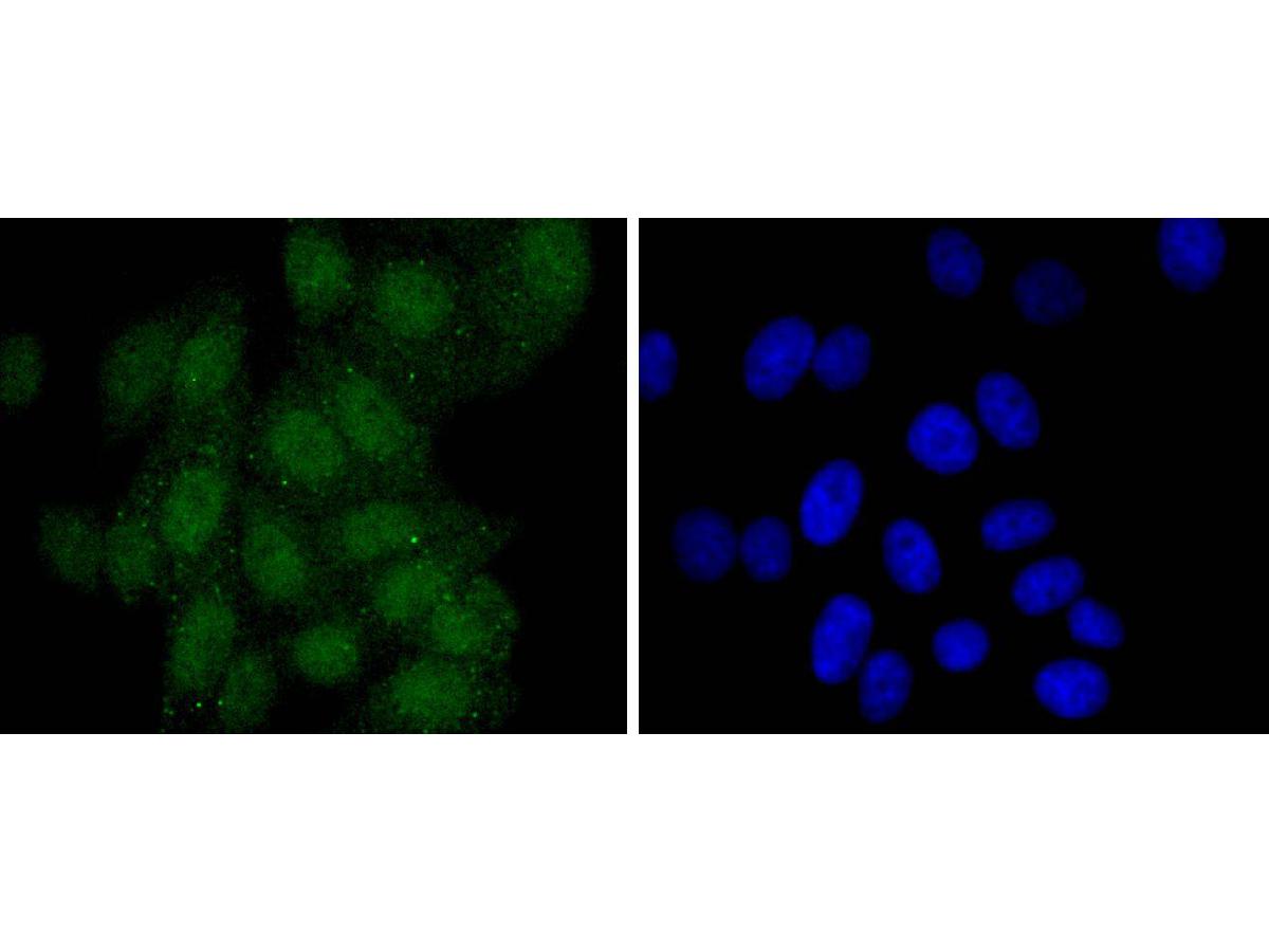
ICC staining of PAX5 in HepG2 cells (green). Formalin fixed cells were permeabilized with 0.1% Triton X-100 in TBS for 10 minutes at room temperature and blocked with 10% negative goat serum for 15 minutes at room temperature. Cells were probed with the primary antibody (ET1701-49, 1/50) for 1 hour at room temperature, washed with PBS. Alexa Fluor®488 conjugate-Goat anti-Rabbit IgG was used as the secondary antibody at 1/1,000 dilution. The nuclear counter stain is DAPI (blue).
-
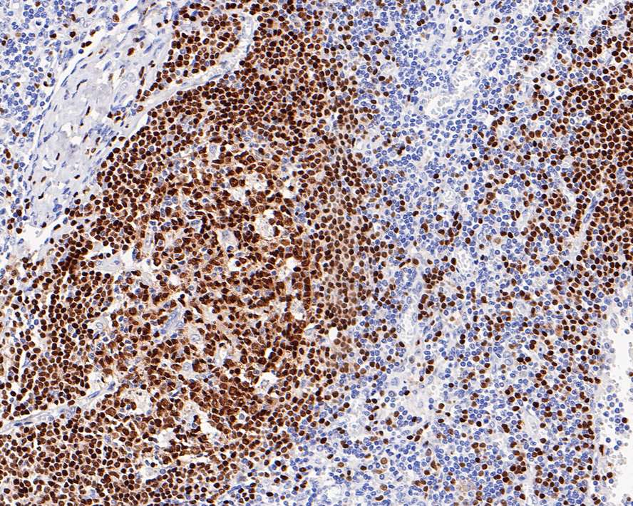
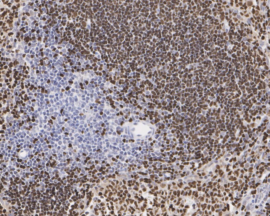
Immunohistochemical analysis of paraffin-embedded human lymph nodes tissue with Rabbit anti-PAX5 antibody (ET1701-49) at 1/1,000 dilution.
The section was pre-treated using heat mediated antigen retrieval with sodium citrate buffer (pH 6.0) for 2 minutes. The tissues were blocked in 1% BSA for 20 minutes at room temperature, washed with ddH2O and PBS, and then probed with the primary antibody (ET1701-49) at 1/1,000 dilution for 1 hour at room temperature. The detection was performed using an HRP conjugated compact polymer system. DAB was used as the chromogen. Tissues were counterstained with hematoxylin and mounted with DPX. -
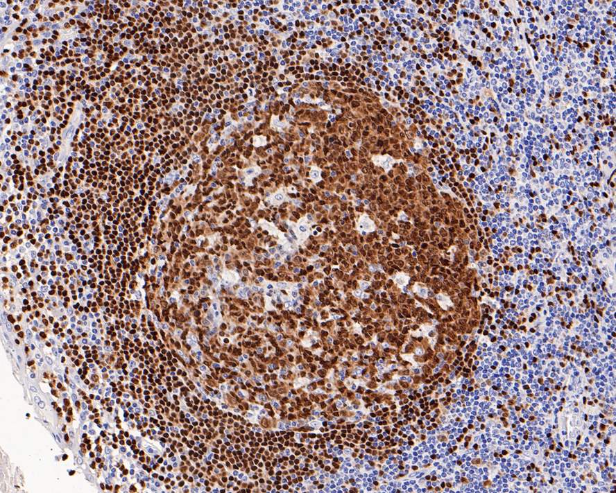
Immunohistochemical analysis of paraffin-embedded human tonsil tissue with Rabbit anti-PAX5 antibody (ET1701-49) at 1/1,000 dilution.
The section was pre-treated using heat mediated antigen retrieval with sodium citrate buffer (pH 6.0) for 2 minutes. The tissues were blocked in 1% BSA for 20 minutes at room temperature, washed with ddH2O and PBS, and then probed with the primary antibody (ET1701-49) at 1/1,000 dilution for 1 hour at room temperature. The detection was performed using an HRP conjugated compact polymer system. DAB was used as the chromogen. Tissues were counterstained with hematoxylin and mounted with DPX. -
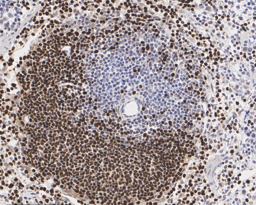
Immunohistochemical analysis of paraffin-embedded human spleen tissue with Rabbit anti-PAX5 antibody (ET1701-49) at 1/1,000 dilution.
The section was pre-treated using heat mediated antigen retrieval with sodium citrate buffer (pH 6.0) for 2 minutes. The tissues were blocked in 1% BSA for 20 minutes at room temperature, washed with ddH2O and PBS, and then probed with the primary antibody (ET1701-49) at 1/1,000 dilution for 1 hour at room temperature. The detection was performed using an HRP conjugated compact polymer system. DAB was used as the chromogen. Tissues were counterstained with hematoxylin and mounted with DPX. -
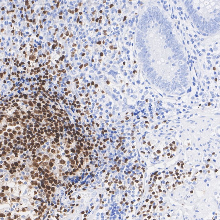
Immunohistochemical analysis of paraffin-embedded mouse spleen tissue with Rabbit anti-PAX5 antibody (ET1701-49) at 1/1,000 dilution.
The section was pre-treated using heat mediated antigen retrieval with sodium citrate buffer (pH 6.0) for 2 minutes. The tissues were blocked in 1% BSA for 20 minutes at room temperature, washed with ddH2O and PBS, and then probed with the primary antibody (ET1701-49) at 1/1,000 dilution for 1 hour at room temperature. The detection was performed using an HRP conjugated compact polymer system. DAB was used as the chromogen. Tissues were counterstained with hematoxylin and mounted with DPX. -
Immunohistochemical analysis of paraffin-embedded rat spleen tissue with Rabbit anti-PAX5 antibody (ET1701-49) at 1/1,000 dilution.
The section was pre-treated using heat mediated antigen retrieval with sodium citrate buffer (pH 6.0) for 2 minutes. The tissues were blocked in 1% BSA for 20 minutes at room temperature, washed with ddH2O and PBS, and then probed with the primary antibody (ET1701-49) at 1/1,000 dilution for 1 hour at room temperature. The detection was performed using an HRP conjugated compact polymer system. DAB was used as the chromogen. Tissues were counterstained with hematoxylin and mounted with DPX. -
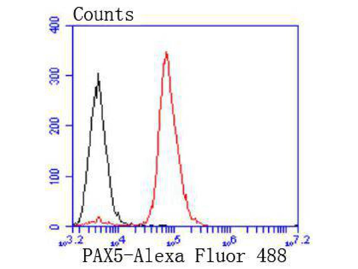
Flow cytometric analysis of PAX5 was done on Raji cells. The cells were fixed, permeabilized and stained with the primary antibody (ET1701-49, 1/50) (red). After incubation of the primary antibody at room temperature for an hour, the cells were stained with a Alexa Fluor®488 conjugate-Goat anti-Rabbit IgG Secondary antibody at 1/1,000 dilution for 30 minutes.Unlabelled sample was used as a control (cells without incubation with primary antibody; black).
Please note: All products are "FOR RESEARCH USE ONLY AND ARE NOT INTENDED FOR DIAGNOSTIC OR THERAPEUTIC USE"