HA tag Recombinant Rabbit Monoclonal Antibody [SN07-06]


Catalog# ET1611-49
HA tag Recombinant Rabbit Monoclonal Antibody [SN07-06]
-
WB
-
IP
-
Species independent
概述
产品名称
HA tag Recombinant Rabbit Monoclonal Antibody [SN07-06]
抗体类型
Recombinant Rabbit monoclonal Antibody
免疫原
Synthetic peptide.
种属反应性
Species independent
验证应用
WB, IP
阳性对照
HA tag recombinant protein, C-terminal HA-tagged recombinant protein.
偶联
unconjugated
克隆号
SN07-06
RRID
产品特性
形态
Liquid
浓度
1ug/ul
存放说明
Store at +4℃ after thawing. Aliquot store at -20℃ or -80℃. Avoid repeated freeze / thaw cycles.
存储缓冲液
1*TBS (pH7.4), 0.05% BSA, 40% Glycerol. Preservative: 0.05% Sodium Azide.
亚型
IgG
纯化方式
Protein A affinity purified.
应用稀释度
-
WB
-
1:1,000-1:5,000
-
IP
-
2-5 µg/ml.
发表文章中的应用
靶点
功能
Human influenza hemagglutinin (HA) is a surface glycoprotein required for the infectivity of the human virus. The HA tag is derived from the HA molecule corresponding to amino acids 98-106. This antibody is used to detect proteins that are tagged with HA tag recombinant protein.
背景文献
1. Liu Z et al. Mark4 promotes oxidative stress and inflammation via binding to PPAR and activating NF-kB pathway in mice adipocytes. Sci Rep 6:21382 (2016).
2. Gauson EJ et al. Evidence supporting a role for TopBP1 and Brd4 in the initiation but not continuation of human papillomavirus 16 E1/E2-mediated DNA replication. J Virol 89:4980-91 (2015).
别名
HA epitope tag antibody
HA1 antibody
HA2 antibody
hemagglutinin antibody
Hemagglutinin HA1 chain antibody
Hemagglutinin HA2 chain antibody
图片
-
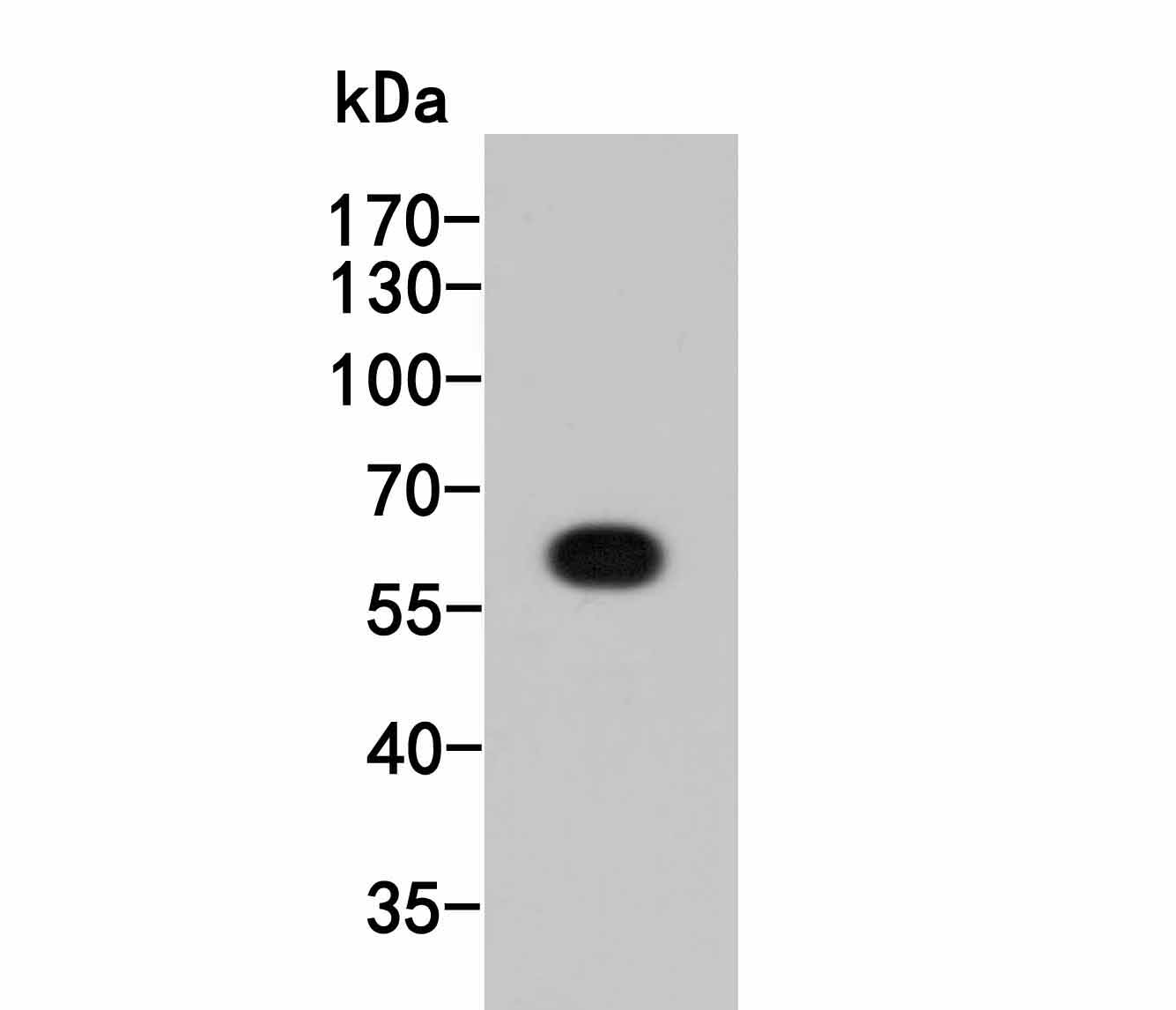
Western blot analysis of HA tag on HA tag recombinant protein. Proteins were transferred to a PVDF membrane and blocked with 5% BSA in PBS for 1 hour at room temperature. The primary antibody (ET1611-49, 1/1,000) was used in 5% BSA at room temperature for 2 hours. Goat Anti-Rabbit IgG - HRP Secondary Antibody (HA1001) at 1:5,000 dilution was used for 1 hour at room temperature.
-
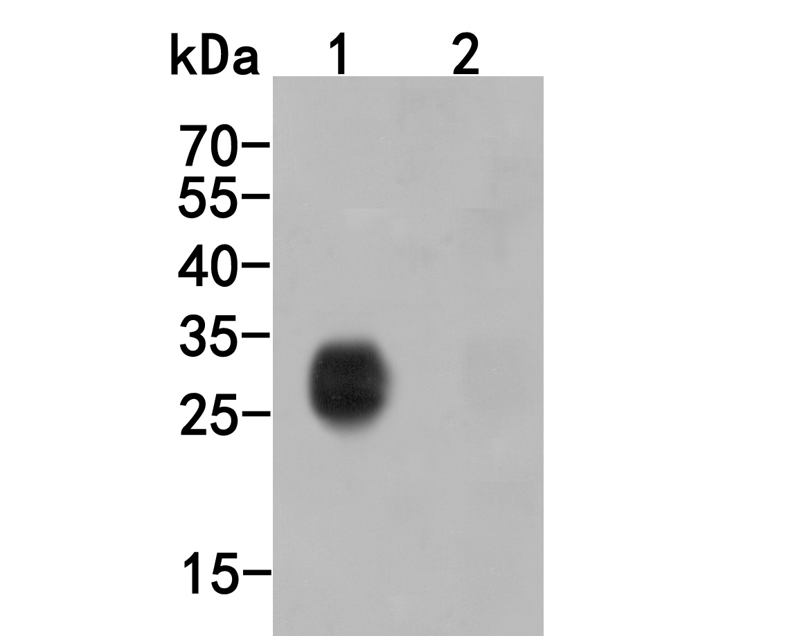
Western blot analysis of HA tag on different lysates. Proteins were transferred to a PVDF membrane and blocked with 5% BSA in PBS for 1 hour at room temperature. The primary antibody (ET1611-49, 1/2,000) was used in 5% BSA at room temperature for 2 hours. Goat Anti-Rabbit IgG - HRP Secondary Antibody (HA1001) at 1:5,000 dilution was used for 1 hour at room temperature.
Positive control:
Lane 1: C-terminal HA-tagged recombinant protein
Lane 2: N-terminal HA-tagged recombinant protein -
HA tag was immunoprecipitated in 5µg C terminal HA Tag fusion protein lysate with ET1611-49 at 2 µg/20 µl agarose. Western blot was performed from the immunoprecipitate using M1008-1 at 1/1,000 dilution. Anti-Mouse IgG - HRP Secondary Antibody (HA1006) at 1:20,000 dilution was used for 60 mins at room temperature.
Lane 1: HA Tag fusion protein lysate (input).
Lane 2: ET1611-49 IP in HA Tag fusion protein lysate.
Lane 3: 0906-1 IP in HA Tag fusion protein lysate.
Lane 4: Rabbit IgG instead of ET1611-49 in HA Tag fusion protein lysate.
Blocking/Dilution buffer: 5% NFDM/TBST
Please note: All products are "FOR RESEARCH USE ONLY AND ARE NOT INTENDED FOR DIAGNOSTIC OR THERAPEUTIC USE"
引文
-
Salmonella Enteritidis T1SS protein SiiD inhibits NLRP3 inflammasome activation via repressing the mtROS-ASC dependent pathway
Author:
PMID: 37155697

应用: WB
反应种属: Mouse
发表时间: 2023 May
-
 Citation
-
The first crystal structure of CD8αα from a cartilaginous fish
Author:
PMID: 37122697

应用: IP
反应种属: Human
发表时间: 2023 Apr
-
Citation
-
Novel Dimeric Architecture of an IFN-γ–Related Cytokine Provides Insights into Subfunctionalization of Type II IFNs in Teleost Fish
Author:
PMID: 36426983

应用: WB
反应种属: Human
发表时间: 2022 Dec
-
Citation
-
Cullin3-TNFAIP1 E3 Ligase Controls Inflammatory Response in Hepatocellular Carcinoma Cells via Ubiquitination of RhoB
Author:
PMID: 33553178

应用: WB
反应种属: Human
发表时间: 2021 Jan
-
Citation
-
Integrative omic analysis reveals the improvement of alkaloid accumulation by ultraviolet-B radiation and its upstream regulation in Catharanthus roseus
Author:
PMID: 33794278

应用: WB
反应种属: Human
发表时间: 2021 Aug
-
Citation
-
The Zscan4-Tet2 Transcription Nexus RegulatesMetabolic Rewiring and Enhances Proteostasis toPromote Reprogramming
Author: Mingliang Zhang;Dan Ye
PMID: 32668244
应用: IP,WB
反应种属: human
发表时间: 2020 Jul
-
Citation
-
Suppression of Th17 cell differentiation by misshapen/NIK-related kinase MINK1.
Author: Linrong Lu
PMID: 28400474
应用: WB
反应种属: mouse
发表时间: 2017 May
-
Citation