Apoptosis Antibody Sampler Kit
RMB: 5300.00
Catalog# K2001
Apoptosis Antibody Sampler Kit
概述
试剂盒组分
| 产品包括 | 规格 | 应用 | 反应性 | MW(kDa) |
|---|---|---|---|---|
| ET1602-39[Caspase-3] | 20µl | WB,IF-Cell,IF-Tissue,IHC-P,IP,FC | Human | Predicted band size: 32 kDa |
| ET1602-47[Active Caspase-3] | 20µl | WB,IF-Cell,IF-Tissue,IHC-P | Human | Predicted band size: 17 kDa |
| ET1608-56[PARP1] | 20µl | WB,IF-Cell,IF-Tissue,IHC-P,FC | Human,Mouse | Predicted band size: 113 kDa |
| ET1608-10[Cleaved PARP] | 20µl | WB,IF-Cell,IP,FC | Human | Predicted band size: 89 kDa |
| ET1610-95[Caspase-9] | 20µl | WB,IP | Human,Mouse | Predicted band size: 46/30/17/37 kDa |
| R1308-12[Active+Pro Caspase-9] | 20µl | WB,IHC-P | Human | Predicted band size: 46 kDa |
| ET1612-28[pro Caspase-7] | 20µl | WB,IF-Cell,IF-Tissue,IHC-P | Human | 34 kDa |
| ER60002[Cleaved-Caspase-7 p20] | 20µl | WB,IF,IHC-P | Human,Mouse,Rat | 20 kDa |
| HA1031[Alpaca anti-Rabbit IgG Fc, Recombinant VHH] | 100µl | IP,ELISA,IHC-P,WB | Rabbit |
产品描述
The Apoptosis Antibody Sampler Kit designed to provide you with a variety of trial-size antibodies in a convenient and cost-effective format. The kit can be used to detect the full length and cleaved products of Caspase-3, Caspase-7, Caspase-9, and PARP. And also includes secondary reagent for detection of these antibodies.
产品特性
存储缓冲液
1*TBS (pH7.4), 0.05% BSA, 40% Glycerol. Preservative: 0.05% Sodium Azide.
存放说明
Store at +4℃ after thawing. Aliquot store at -20℃. Avoid repeated freeze / thaw cycles.
背景
Apoptosis is a regulated physiological process leading to cell death. Caspases, a family of cysteine acid proteases, are central regulators of apoptosis. Initiator caspases are closely coupled to proapoptotic signals. </br>Once activated, these caspases cleave and activate downstream effector caspases, which in turn cleave cytoskeletal and nuclear proteins like PARP, α-fodrin, DFF, and lamin A and induce apoptosis. Cytochrome c released from mitochondria is coupled to the activation of caspase-9, a key initiator caspase. Proapoptotic stimuli include FasL, TNF-α, DNA damage and ER stress. Fas and TNFR activate caspase-8 and -10, DNA damage leads to the activation of caspase-9 and ER stress leads to the calcium-mediated activation of caspase-12 (3). The inhibitor of apoptosis protein (IAP) family includes XIAP and survivin and functions by binding and inhibiting several caspases. Smac/Diablo, a mitochondrial protein, is released into the cytosol upon mitochondrial stress and competes with caspases for binding of IAPs. The interaction of Smac/Diablo with IAPs relieves the inhibitory effects of IAPs on caspases .
背景文献
2. Budihardjo, I. et al. (1999) Annu. Rev. Cell Dev. Biol. 15, 269-290.
3. Nakagawa, T. et al. (2000) Nature 403, 98-103.
4. Deveraux, Q. L. et al. (1998) EMBO J. 17, 2215-2223.
5. Li, F. et al. (1998) Nature 396, 580-584.
6. Du, C. et al. (2000) Cell 102, 33-42.
图片
-
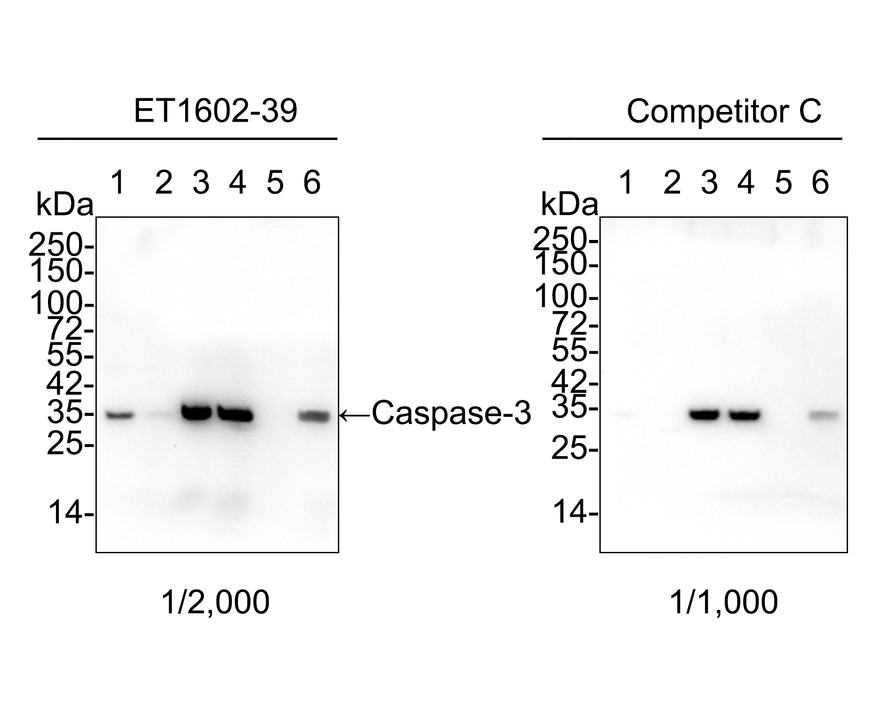
Western blot analysis of Caspase-3 on different lysates with Rabbit anti-Caspase-3 antibody (ET1602-39) at 1/2,000 dilution and competitor's antibody at 1/1,000 dilution.
Lane 1: HeLa cell lysate
Lane 2: HeLa treated with 1μM staurosporine for 3 hours cell lysate
Lane 3: Jurkat cell lysate
Lane 4: Jurkat treated with 25μM Etoposide for 5 hours cell lysate
Lane 5: MCF7 cell lysate (negative)
Lane 6: HEK-293 cell lysate
Lysates/proteins at 20 µg/Lane.
Predicted band size: 32 kDa
Observed band size: 32 kDa
Exposure time: 3 minutes 20 seconds;
4-20% SDS-PAGE gel.
Proteins were transferred to a PVDF membrane and blocked with 5% NFDM/TBST for 1 hour at room temperature. The primary antibody (ET1602-39) at 1/2,000 dilution and competitor's antibody at 1/1,000 dilution were used in 5% NFDM/TBST at 4℃ overnight. Goat Anti-Rabbit IgG - HRP Secondary Antibody (HA1001) at 1/50,000 dilution was used for 1 hour at room temperature. -
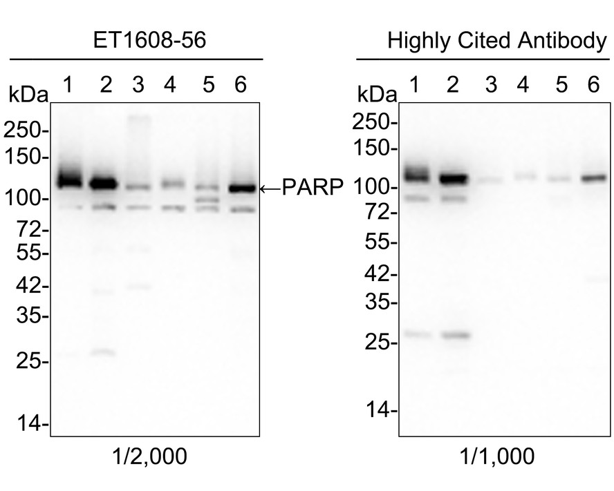
Western blot analysis of PARP on different lysates with Rabbit anti-PARP antibody (ET1608-56) at 1/2,000 dilution and competitor's antibody at 1/1,000 dilution.
Lane 1: HeLa cell lysate (15 µg/Lane)
Lane 2: Jurkat cell lysate (15 µg/Lane)
Lane 3: NIH/3T3 cell lysate (15 µg/Lane)
Lane 4: C2C12 cell lysate (15 µg/Lane)
Lane 5: C6 cell lysate (15 µg/Lane)
Lane 6: PC-12 cell lysate (15 µg/Lane)
Predicted band size: 113 kDa
Observed band size: 113 kDa
Exposure time: 6 seconds;
4-20% SDS-PAGE gel.
Proteins were transferred to a PVDF membrane and blocked with 5% NFDM/TBST for 1 hour at room temperature. The primary antibody (ET1608-56) at 1/2,000 dilution and competitor's antibody at 1/1,000 dilution were used in 5% NFDM/TBST at 4℃ overnight. Goat Anti-Rabbit IgG - HRP Secondary Antibody (HA1001) at 1:50,000 dilution was used for 1 hour at room temperature. -
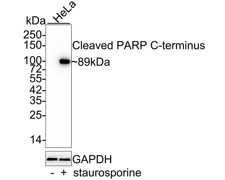
Western blot analysis of Cleaved PARP on different lysates with Rabbit anti-Cleaved PARP antibody (ET1608-10) at 1/2,000 dilution.
Lane 1: HeLa whole cell lysate
Lane 2: HeLa treated with 1μM staurosporine for 3 hours whole cell lysate
Lysates/proteins at 20 µg/Lane.
Predicted band size: 89 kDa
Observed band size: 89 kDa
Exposure time: 1 minute 9 seconds;
4-20% SDS-PAGE gel.
Proteins were transferred to a PVDF membrane and blocked with 5% NFDM/TBST for 1 hour at room temperature. The primary antibody (ET1608-10) at 1/2,000 dilution was used in 5% NFDM/TBST at 4℃ overnight. Goat Anti-Rabbit IgG - HRP Secondary Antibody (HA1001) at 1:50,000 dilution was used for 1 hour at room temperature. -
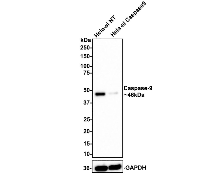
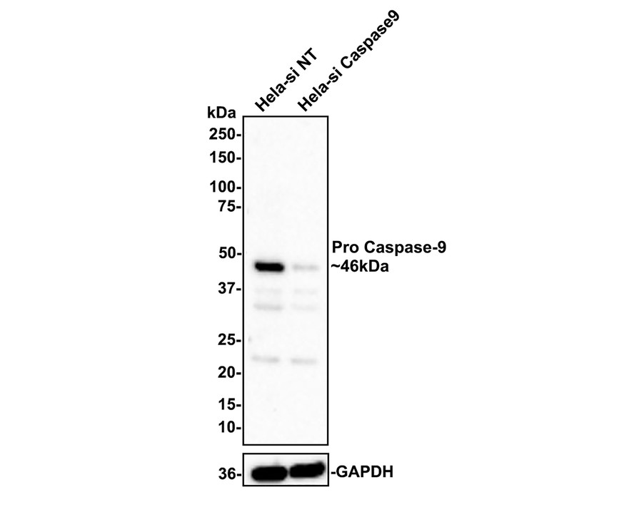
Western blot analysis of Caspase-9 on different lysates with Rabbit anti-Caspase-9 antibody (ET1610-95) at 1/1,000 dilution.
Lane 1: Hela-si NT cell lysate
Lane 2: Hela-si Caspase-9 cell lysate
Lysates/proteins at 10 µg/Lane.
Predicted band size: 46 kDa
Observed band size: 46 kDa
Exposure time: 2 minutes;
4-20% SDS-PAGE gel.
ET1610-95 was shown to specifically react with Caspase-9 in Hela-si NT cells. Weakened band was observed when Hela-si Caspase-9 sample was tested. Hela-si NT and Hela-si Caspase-9 samples were subjected to SDS-PAGE. Proteins were transferred to a PVDF membrane and blocked with 5% NFDM in TBST for 1 hour at room temperature. The primary antibody (ET1610-95, 1/1,000) and Loading control antibody (Rabbit anti-GAPDH, ET1601-4, 1/10,000) were used in 5% BSA at room temperature for 2 hours. Goat Anti-rabbit IgG-HRP Secondary Antibody (HA1001) at 1:100,000 dilution was used for 1 hour at room temperature. -
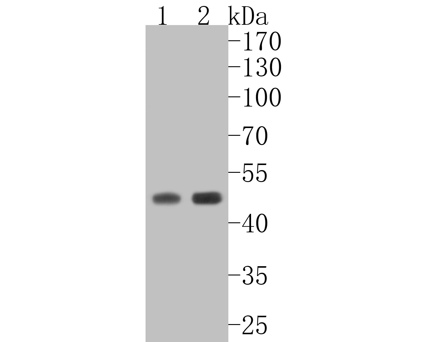
Western blot analysis of Active+Pro Caspase-9 on different lysates with Rabbit anti-Active+Pro Caspase-9 antibody (R1308-12) at 1/1,000 dilution.
Lane 1: Hela-si NT cell lysate
Lane 2: Hela-si Caspase-9 cell lysate
Lysates/proteins at 10 µg/Lane.
Predicted band size: 46 kDa
Observed band size: 46 kDa
Exposure time: 5 minutes;
4-20% SDS-PAGE gel.
R1308-12 was shown to specifically react with Active+Pro Caspase-9 in Hela-si NT cells. Weakened band was observed when Hela-si Active+Pro Caspase-9 sample was tested. Hela-si NT and Hela-si Active+Pro Caspase-9 samples were subjected to SDS-PAGE. Proteins were transferred to a PVDF membrane and blocked with 5% NFDM in TBST for 1 hour at room temperature. The primary antibody (R1308-12, 1/1,000) and Loading control antibody (Rabbit anti-GAPDH, ET1601-4, 1/10,000) were used in 5% BSA at room temperature for 2 hours. Goat Anti-rabbit IgG-HRP Secondary Antibody (HA1001) at 1:100,000 dilution was used for 1 hour at room temperature.
Related Products
Caspase-9 Recombinant Rabbit Monoclonal Antibody [SZ29-01]
Application: WB,IF-Cell,IF-Tissue,IHC-P,IP,FC
Reactivity: Human,Mouse
Conjugate: unconjugated

Active+Pro Caspase-9 Rabbit Polyclonal Antibody
Application: WB,IHC-P
Reactivity: Human
Conjugate: unconjugated
Caspase-9 Recombinant Rabbit Monoclonal Antibody [SC65-05]
Application: WB,IP
Reactivity: Human,Mouse
Conjugate: unconjugated
